Copyright
©The Author(s) 2024.
World J Gastroenterol. Feb 28, 2024; 30(8): 817-832
Published online Feb 28, 2024. doi: 10.3748/wjg.v30.i8.817
Published online Feb 28, 2024. doi: 10.3748/wjg.v30.i8.817
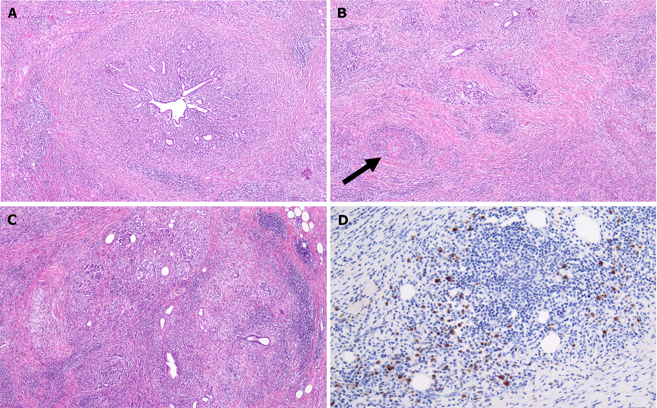
Figure 5 Histology of autoimmune pancreatitis.
A: Histological samples of type 1 autoimmune pancreatitis. Hematoxylin eosin (HE) 4, Duct centric lymphoplasmacytic infiltrate; B: HE 10, storiform fibrosis with intense lymphoplasmacytic infiltrate and obliterative phlebitis (arrow); C: HE 4, lobule complete effacement by inflammatory cells and fibrosis; D: Immunoglobulin G4 (IgG4) IIC 20, moderate increase of IgG4+ plasma cells.
- Citation: Gallo C, Dispinzieri G, Zucchini N, Invernizzi P, Massironi S. Autoimmune pancreatitis: Cornerstones and future perspectives. World J Gastroenterol 2024; 30(8): 817-832
- URL: https://www.wjgnet.com/1007-9327/full/v30/i8/817.htm
- DOI: https://dx.doi.org/10.3748/wjg.v30.i8.817


