Copyright
©The Author(s) 2024.
World J Gastroenterol. Feb 28, 2024; 30(8): 817-832
Published online Feb 28, 2024. doi: 10.3748/wjg.v30.i8.817
Published online Feb 28, 2024. doi: 10.3748/wjg.v30.i8.817
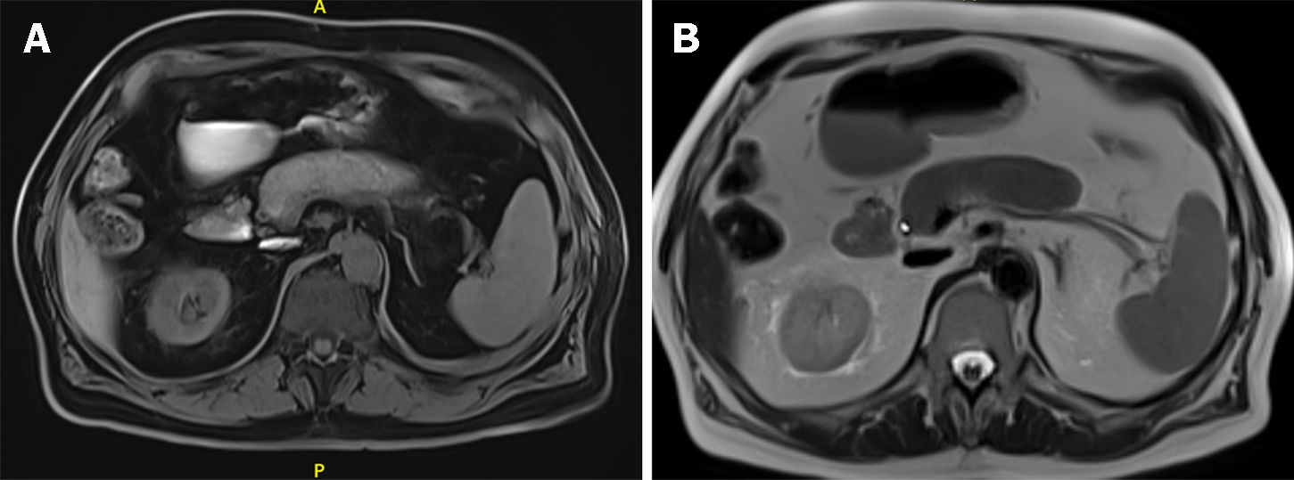
Figure 3 Radiological appearance of autoimmune pancreatitis-part 2.
A: Unenhanced T1 weighted magnetic resonance imaging (MRI) images of autoimmune pancreatitis (AIP): Diffuse hypointense pancreas, with an even more hypointense fibrotic capsule-rim; B: Unenhanced T2 weighted MRI images of diffuse AIP: The affected parenchyma shows a moderately higher intensity signal, with a persistently low-intensity fibrotic rim.
- Citation: Gallo C, Dispinzieri G, Zucchini N, Invernizzi P, Massironi S. Autoimmune pancreatitis: Cornerstones and future perspectives. World J Gastroenterol 2024; 30(8): 817-832
- URL: https://www.wjgnet.com/1007-9327/full/v30/i8/817.htm
- DOI: https://dx.doi.org/10.3748/wjg.v30.i8.817


