Copyright
©The Author(s) 2024.
World J Gastroenterol. Feb 21, 2024; 30(7): 705-713
Published online Feb 21, 2024. doi: 10.3748/wjg.v30.i7.705
Published online Feb 21, 2024. doi: 10.3748/wjg.v30.i7.705
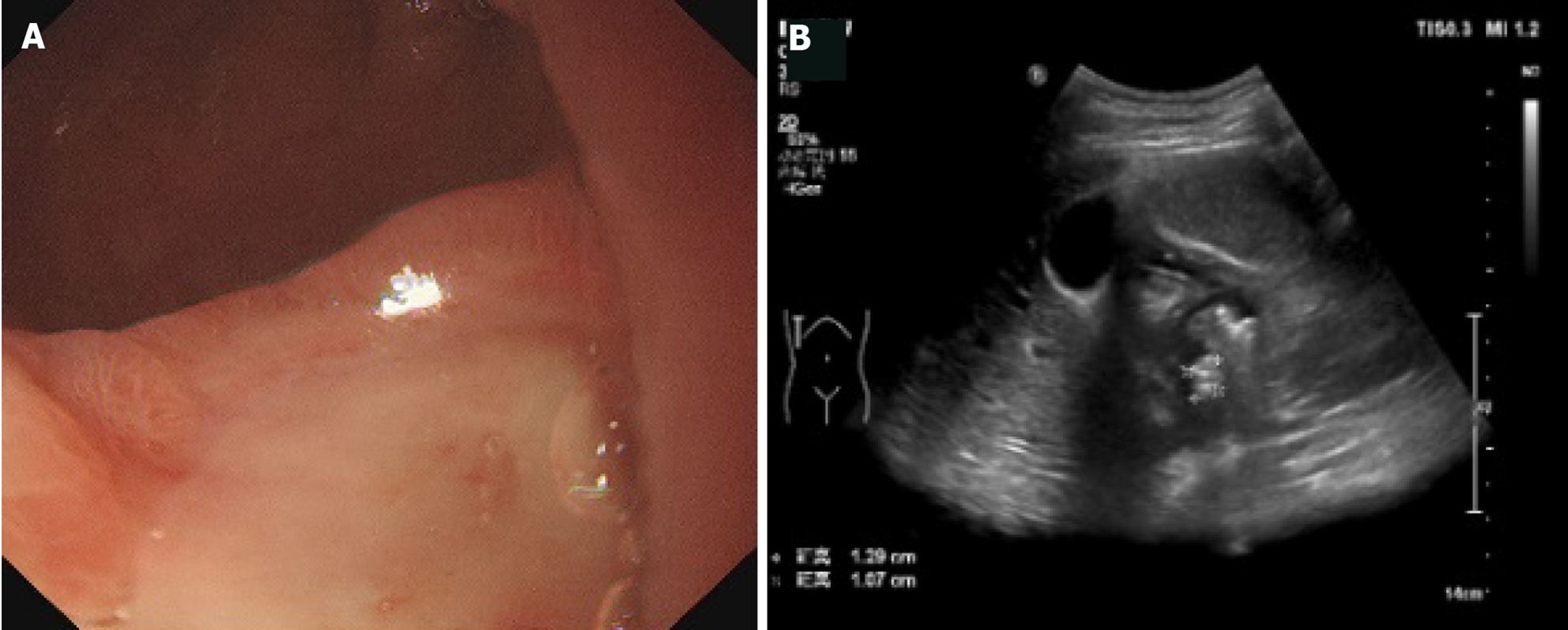
Figure 2 A 14-year-old male with duodenal ulcer diagnosed by gastroscopy.
A: Gastroscope indicated a large ulcer on the anterior wall of the bulb, covered with thick white moss, congestion and edema of the surrounding mucosa; B: Contrast-enhanced ultrasonography showed that the shape of duodenal bulb was irregular, the area was small, and there were hyperechoic plaques, 13 mm × 11 mm × 13 mm in size, on the anterior wall of the duodenum.
- Citation: Zhang YH, Xu ZH, Ni SS, Luo HX. Gastrointestinal contrast-enhanced ultrasonography for diagnosis and treatment of peptic ulcer in children. World J Gastroenterol 2024; 30(7): 705-713
- URL: https://www.wjgnet.com/1007-9327/full/v30/i7/705.htm
- DOI: https://dx.doi.org/10.3748/wjg.v30.i7.705


