Copyright
©The Author(s) 2024.
World J Gastroenterol. Feb 14, 2024; 30(6): 542-555
Published online Feb 14, 2024. doi: 10.3748/wjg.v30.i6.542
Published online Feb 14, 2024. doi: 10.3748/wjg.v30.i6.542
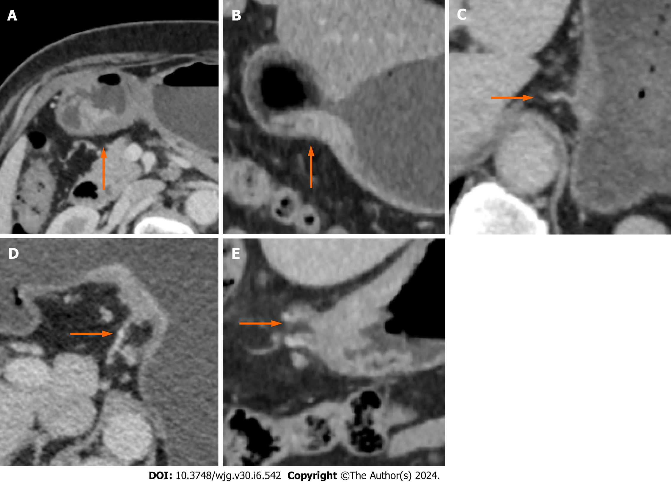
Figure 2 Example of a computed tomography-detected extramural vein invasion score on computed tomography images of gastric cancer patients.
A: Score 0: The tumor has not penetrated the gastric wall, and there are no extramural vessels beside the lesion (arrow) in the transverse position of the venous phase (VP); B: Score 1: The transverse view of the VP shows that the tumor has permeated the gastric wall, and there are no extramural vessels beside the lesion (arrow); C: Score 2: In the VP, the coronal lesion has penetrated the gastric wall, and there are tortuous blood vessels connected with the lesion (arrow), but no tumor density shadow is observed in the vascular lumen; D: Score 3: The transverse view of the VP shows that the mass has penetrated through the gastric wall, the involved blood vessels appear slightly tortuous and dilated, and the tumor density shadow is visible (arrow); E: Score 4: In the coronary view of the VP, the tumor permeated the gastric wall, the extramural vascular lumen was significantly dilated, and a slight low-density filling defect was visible inside (arrow).
- Citation: Ge HT, Chen JW, Wang LL, Zou TX, Zheng B, Liu YF, Xue YJ, Lin WW. Preoperative prediction of lymphovascular and perineural invasion in gastric cancer using spectral computed tomography imaging and machine learning. World J Gastroenterol 2024; 30(6): 542-555
- URL: https://www.wjgnet.com/1007-9327/full/v30/i6/542.htm
- DOI: https://dx.doi.org/10.3748/wjg.v30.i6.542


