Copyright
©The Author(s) 2024.
World J Gastroenterol. Dec 28, 2024; 30(48): 5111-5129
Published online Dec 28, 2024. doi: 10.3748/wjg.v30.i48.5111
Published online Dec 28, 2024. doi: 10.3748/wjg.v30.i48.5111
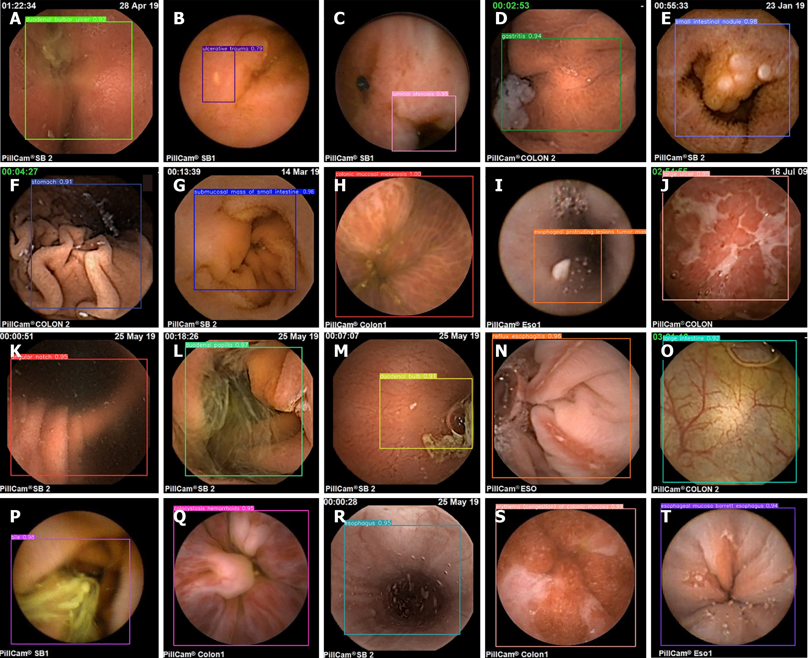
Figure 11 Wireless capsule endoscopy_detection model detection visualization results.
Different letters in the image represent different types of lesions. A: Duodenal bulbar ulcer; B: Ulcerative trauma; C: Luminal stenosis; D: Gastritis; E: Small intestinal nodule; F: Stomach; G: Submucosal mass of the small intestine; H: Colonic mucosal melanosis; I: Esophageal protruding lesion tumor mass; J: Large ulcer; K: Angular notch; L: Duodenal papilla; M: Duodenal bulb; N: Reflux esophagitis; O: Large intestine; P: Bile; Q: Colocystosis hemorrhoids; R: Esophagus; S: Gastric antrum; T: Duodenal bulbar erosion.
- Citation: Xiao ZG, Chen XQ, Zhang D, Li XY, Dai WX, Liang WH. Image detection method for multi-category lesions in wireless capsule endoscopy based on deep learning models. World J Gastroenterol 2024; 30(48): 5111-5129
- URL: https://www.wjgnet.com/1007-9327/full/v30/i48/5111.htm
- DOI: https://dx.doi.org/10.3748/wjg.v30.i48.5111


