Copyright
©The Author(s) 2024.
World J Gastroenterol. Dec 14, 2024; 30(46): 4937-4946
Published online Dec 14, 2024. doi: 10.3748/wjg.v30.i46.4937
Published online Dec 14, 2024. doi: 10.3748/wjg.v30.i46.4937
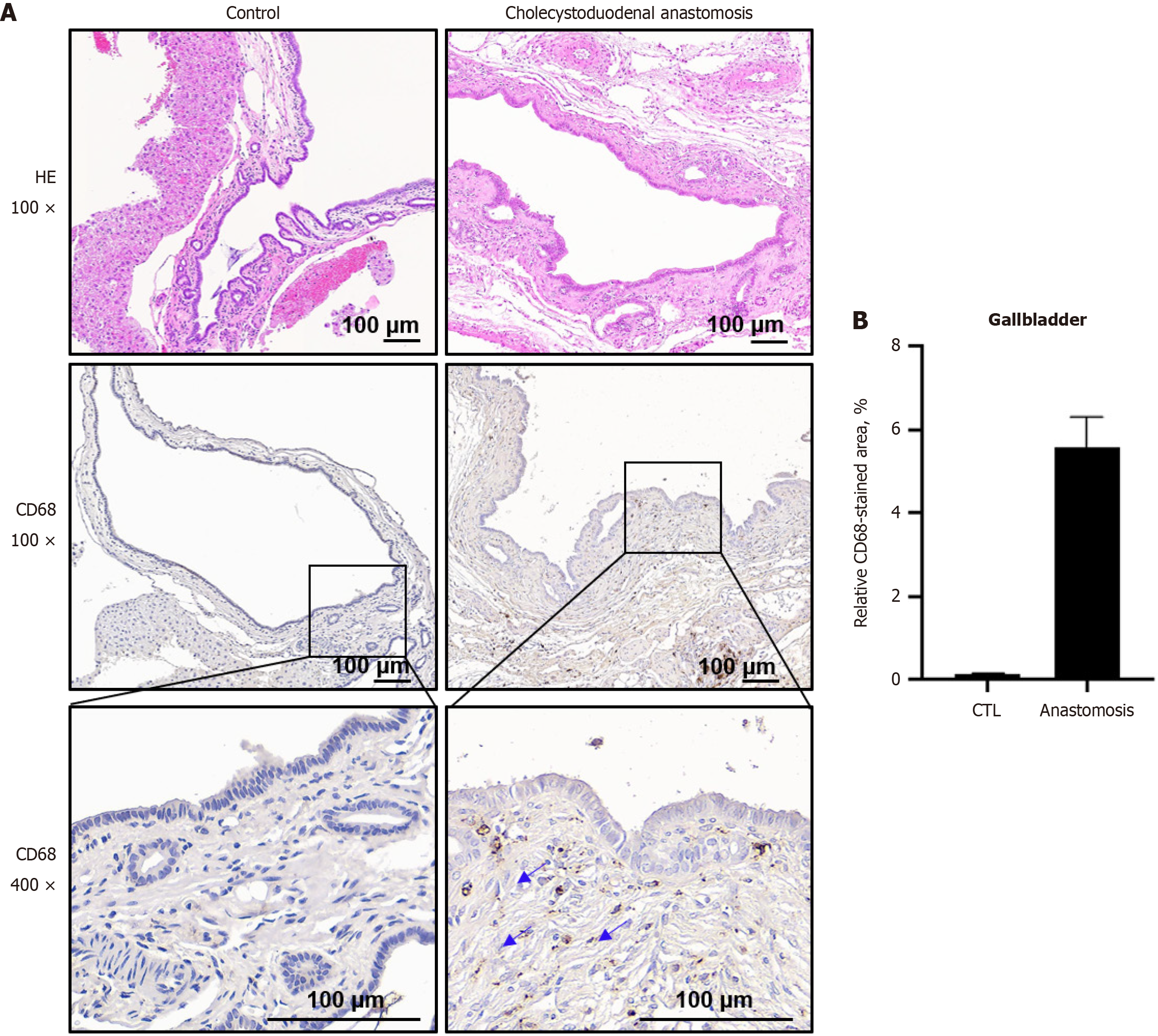
Figure 4 Structural changes and inflammation in the gallbladder epithelium of control and cholecystoduodenal anastomosis mice.
A: Upper panel: Hematoxylin and eosin staining of the gallbladder epithelium from control and cholecystoduodenal anastomosis mice. Lower panel: Immunohistochemical staining with CD68 in gallbladder tissues from control mice (left) and cholecystoduodenal anastomosis mice (right); B: Relative CD68-stained area was represented as the mean ± SD. CTL: Control.
- Citation: Jang Y, Kim JY, Han SY, Park A, Baek SJ, Lee G, Kang J, Ryu H, Kim SH. Establishment of a chronic biliary disease mouse model with cholecystoduodenal anastomosis for intestinal microbiome preservation. World J Gastroenterol 2024; 30(46): 4937-4946
- URL: https://www.wjgnet.com/1007-9327/full/v30/i46/4937.htm
- DOI: https://dx.doi.org/10.3748/wjg.v30.i46.4937


