Copyright
©The Author(s) 2024.
World J Gastroenterol. Dec 7, 2024; 30(45): 4817-4835
Published online Dec 7, 2024. doi: 10.3748/wjg.v30.i45.4817
Published online Dec 7, 2024. doi: 10.3748/wjg.v30.i45.4817
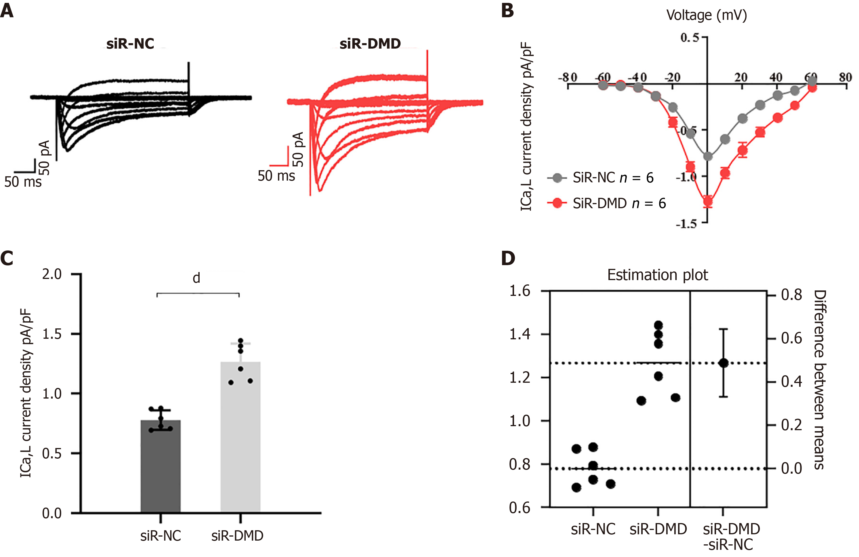
Figure 5 Downregulation of dystrophin expression leads to dysregulation of calcium influx in human intestinal smooth muscle cells.
A: Representative traces of L-type calcium currents (ICa, L) were recorded from human intestinal smooth muscle cells transfected with negative control small interfering RNA (siR-NC) or siR-dystrophin (siR-DMD) using patch-clamp techniques (the X-axis represents the time in milliseconds and the Y-axis represents the current magnitude in pico-amps); B: Line graph of ICa-L current density. The inward current induced by depolarization voltage increased after siR-DMD transfection; C: Peak intracellular calcium concentrations in the two groups of human intestinal smooth muscle cells; D: Average difference between cells transfected with siR-NC or siR-DMD shown in the estimated graph above. Both groups are plotted on the left Y-axis; the mean difference is shown as a bootstrap sampling distribution on the floating axis on the right. dP < 0.0001, n = 6. siR-NC: Negative control small interfering RNA; siR-DMD: Dystrophin small interfering RNA; DMD: Dystrophin; ICa-L: L-type calcium currents.
- Citation: Li WZ, Xiong Y, Wang TK, Chen YY, Wan SL, Li LY, Xu M, Tong JJ, Qian Q, Jiang CQ, Liu WC. Quantitative proteomics analysis reveals the pathogenesis of obstructed defecation syndrome caused by abnormal expression of dystrophin. World J Gastroenterol 2024; 30(45): 4817-4835
- URL: https://www.wjgnet.com/1007-9327/full/v30/i45/4817.htm
- DOI: https://dx.doi.org/10.3748/wjg.v30.i45.4817


