Copyright
©The Author(s) 2024.
World J Gastroenterol. Jan 28, 2024; 30(4): 367-380
Published online Jan 28, 2024. doi: 10.3748/wjg.v30.i4.367
Published online Jan 28, 2024. doi: 10.3748/wjg.v30.i4.367
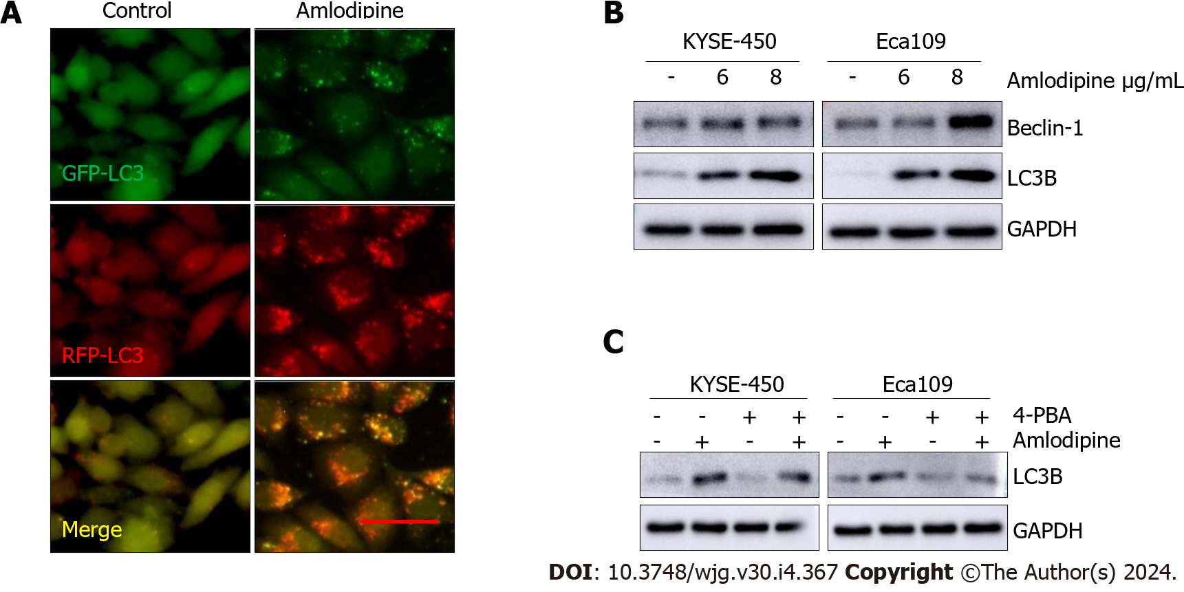
Figure 5 Amlodipine induced autophagy in esophageal carcinoma cells.
A: Eca109 cells after transient infection with GFP-RFP-LC3 virus were treated with vehicle control PBS or amlodipine (6 μg/mL) for 24 h and representative fluorescence images were captured. Bar = 20 μm; B: Protein trajectory results were measured using PBS or different concentrations of amlodipine, and the levels of beclin-1 and light chain 3 B (LC3B) in KYSE-450 and Eca109 cells after two days of treatment were detected; C: Western blot analysis revealed the level of LC3B in KYSE-450 and Eca109 after 48 h treatment with amlodipine (4 μg/mL), 4-phenylbutyric acid (4-PBA) (0.2 μM) or both combination (applying 4-PBA in advance for 1 h then amlodipine). GAPDH served as a loading control.
- Citation: Chen YM, Yang WQ, Gu CW, Fan YY, Liu YZ, Zhao BS. Amlodipine inhibits the proliferation and migration of esophageal carcinoma cells through the induction of endoplasmic reticulum stress. World J Gastroenterol 2024; 30(4): 367-380
- URL: https://www.wjgnet.com/1007-9327/full/v30/i4/367.htm
- DOI: https://dx.doi.org/10.3748/wjg.v30.i4.367


