Copyright
©The Author(s) 2024.
World J Gastroenterol. Jan 28, 2024; 30(4): 367-380
Published online Jan 28, 2024. doi: 10.3748/wjg.v30.i4.367
Published online Jan 28, 2024. doi: 10.3748/wjg.v30.i4.367
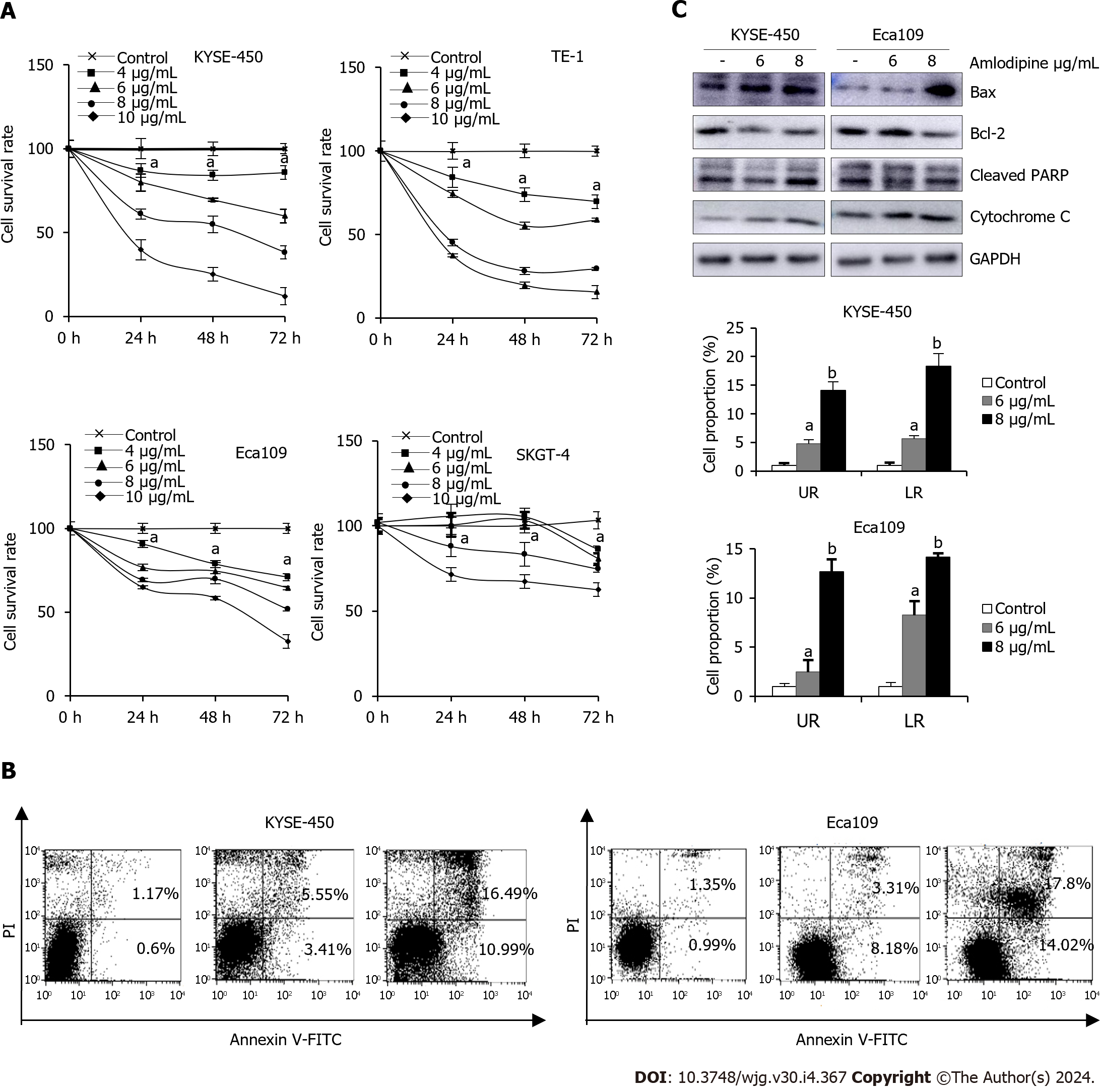
Figure 2 Amlodipine suppressed esophageal carcinoma cell growth through mitochondria-mediated apoptosis.
A: For human esophageal cancer cells, treatment with different concentrations of amlodipine solution at 4-10 μg/mL, increasing by 2 μg/mL, was performed. Cell viability was measured using the 3-(4,5-dimethyl-2-thiazolyl)-2,5-diphenyl tetrazolium bromide (MTT) assay to observe the state of cells after treatment for one, two, and three days aP < 0.05 vs Control; B: KYSE-450 and Eca109 cells were exposed to amlodipine at a concentration of either 6 or 8 μg/mL for 48 h, following which the percentage of apoptotic cells was measured using flow cytometry or propidium iodide staining aP < 0.05 vs Control, bP < 0.01 vs Control; C: In addition, after two days of treatment with amlodipine, changes in the levels of various apoptosis-related proteins including Bcl-2, Bax, cleaved PARP, and cytochrome C were detected using Western blotting techniques. UR: Upper right quadrant LR: Lower right quadrant.
- Citation: Chen YM, Yang WQ, Gu CW, Fan YY, Liu YZ, Zhao BS. Amlodipine inhibits the proliferation and migration of esophageal carcinoma cells through the induction of endoplasmic reticulum stress. World J Gastroenterol 2024; 30(4): 367-380
- URL: https://www.wjgnet.com/1007-9327/full/v30/i4/367.htm
- DOI: https://dx.doi.org/10.3748/wjg.v30.i4.367


