Copyright
©The Author(s) 2024.
World J Gastroenterol. Oct 14, 2024; 30(38): 4232-4238
Published online Oct 14, 2024. doi: 10.3748/wjg.v30.i38.4232
Published online Oct 14, 2024. doi: 10.3748/wjg.v30.i38.4232
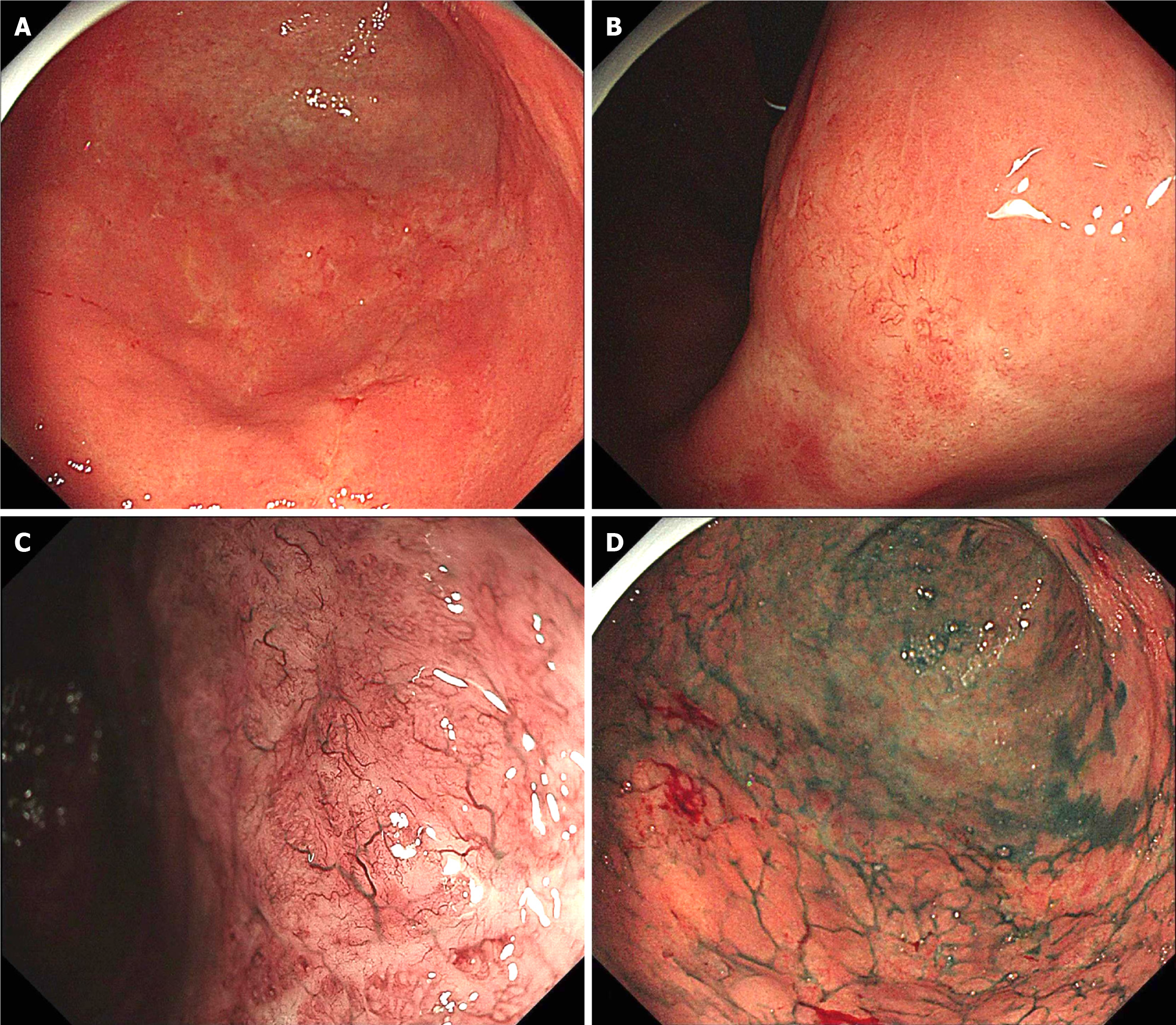
Figure 1 Endoscopic features of lesions.
A: Whitish, shallow, and uneven mucosa lesions with minimal spontaneous bleeding at gastric antrum; B: The tumor invades gastric antrum and angular notch; C: Magnifying endoscopy with narrow-band imaging reveals typical tree-like appearance microvessels; D: Indigo carmine chromoendoscopy delineates a poor-demarcated lesion with an irregular margin.
- Citation: Jia YF, Chen FF, Yang L, Ye YX, Gao YZ, Zhang WY, Yang JL. Early gastric composite tumor comprising signet-ring cell carcinoma and mucosa-associated lymphoid tissue lymphoma: A case report. World J Gastroenterol 2024; 30(38): 4232-4238
- URL: https://www.wjgnet.com/1007-9327/full/v30/i38/4232.htm
- DOI: https://dx.doi.org/10.3748/wjg.v30.i38.4232


