Copyright
©The Author(s) 2024.
World J Gastroenterol. Oct 7, 2024; 30(37): 4115-4131
Published online Oct 7, 2024. doi: 10.3748/wjg.v30.i37.4115
Published online Oct 7, 2024. doi: 10.3748/wjg.v30.i37.4115
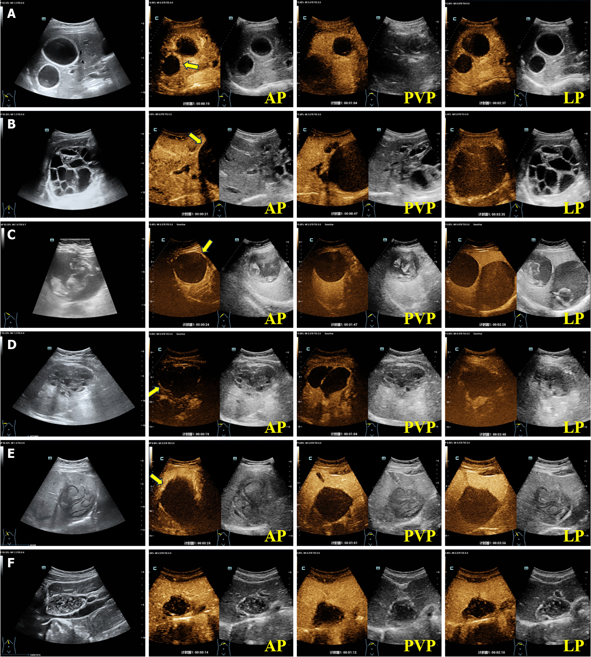
Figure 3 Contrast-enhanced ultrasound images of hepatic cystic echinococcosis.
A: A 27-year-old female patient presenting with a liver lesion of hepatic cystic echinococcosis 1 (HCE1), exhibiting peripheral “rim-like” enhancement in the arterial phase (AP) on contrast-enhanced ultrasound (CEUS) (the yellow arrow), with no internal enhancement observed; B: A 41-year-old female patient presenting with a liver lesion of HCE2, exhibiting slightly peripheral “rim-like” enhancement in the AP on CEUS (the yellow arrow), with no internal enhancement detected; C: A 56-year-old female patient presenting with a liver lesion of HCE3a, exhibiting slightly peripheral “rim-like” enhancement in the AP on CEUS (the yellow arrow), with no internal enhancement observed; D: A 49-year-old male patient presenting with a liver lesion of HCE3b, exhibiting slightly peripheral “rim-like” enhancement in the AP on CEUS (the yellow arrow), with internal septal enhancement visible; E: A 47-year-old female patient presenting with a liver lesion of HCE4, exhibiting slightly peripheral “rim-like” enhancement in the AP on CEUS (the yellow arrow), with no internal enhancement observed; F: A 29-year-old female patient presenting with a liver lesion of HCE5, with no obvious enhancement in the AP, portal venous phase, or late phase.
- Citation: Tao Y, Wang YF, Wang J, Long S, Seyler BC, Zhong XF, Lu Q. Pictorial review of hepatic echinococcosis: Ultrasound imaging and differential diagnosis. World J Gastroenterol 2024; 30(37): 4115-4131
- URL: https://www.wjgnet.com/1007-9327/full/v30/i37/4115.htm
- DOI: https://dx.doi.org/10.3748/wjg.v30.i37.4115


