Copyright
©The Author(s) 2024.
World J Gastroenterol. Oct 7, 2024; 30(37): 4115-4131
Published online Oct 7, 2024. doi: 10.3748/wjg.v30.i37.4115
Published online Oct 7, 2024. doi: 10.3748/wjg.v30.i37.4115
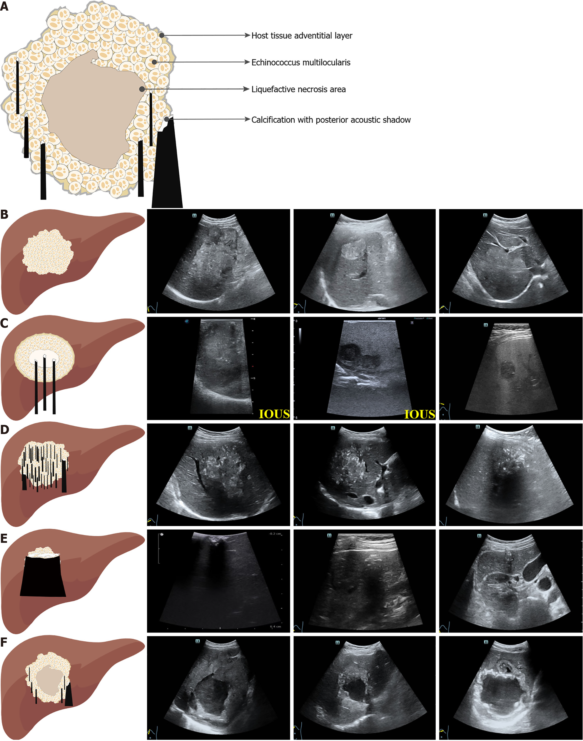
Figure 2 Schematic diagrams and grayscale ultrasound images of hepatic alveolar echinococcosis.
A: Schematic structure of hepatic alveolar echinococcosis (HAE); B: Hemangioma-like: Hemangioma-like lesions of HAE manifest as well-defined and heterogeneous hyperechoic masses in comparison to the surrounding hepatic parenchyma; C: Metastasis-like: Metastasis-like lesions of HAE manifest as hypoechoic masses with indistinct margins, lacking a halo sign, and with a hyperechoic and heterogeneous scar at their center; D: Hailstorm: Hailstorm lesions of HAE are characterized by ill-defined boundaries and irregularly shaped masses of heterogeneous echogenicity, accompanied by scattered or diffuse calcifications, with or without a posterior acoustic shadow; E: Ossification: Ossified lesions of HAE present as small and sharply delineated lesions with internal clumps of calcification and a posterior acoustic shadow; F: Pseudocystic: Pseudocystic lesions of HAE are primarily characterized by a thickness exceeding 10 mm with high echogenicity, irregular and non-uniform margins, and a low-echogenicity liquefied necrotic area inside. IOUS: Intraoperative ultrasound.
- Citation: Tao Y, Wang YF, Wang J, Long S, Seyler BC, Zhong XF, Lu Q. Pictorial review of hepatic echinococcosis: Ultrasound imaging and differential diagnosis. World J Gastroenterol 2024; 30(37): 4115-4131
- URL: https://www.wjgnet.com/1007-9327/full/v30/i37/4115.htm
- DOI: https://dx.doi.org/10.3748/wjg.v30.i37.4115


