Copyright
©The Author(s) 2024.
World J Gastroenterol. Sep 28, 2024; 30(36): 4071-4077
Published online Sep 28, 2024. doi: 10.3748/wjg.v30.i36.4071
Published online Sep 28, 2024. doi: 10.3748/wjg.v30.i36.4071
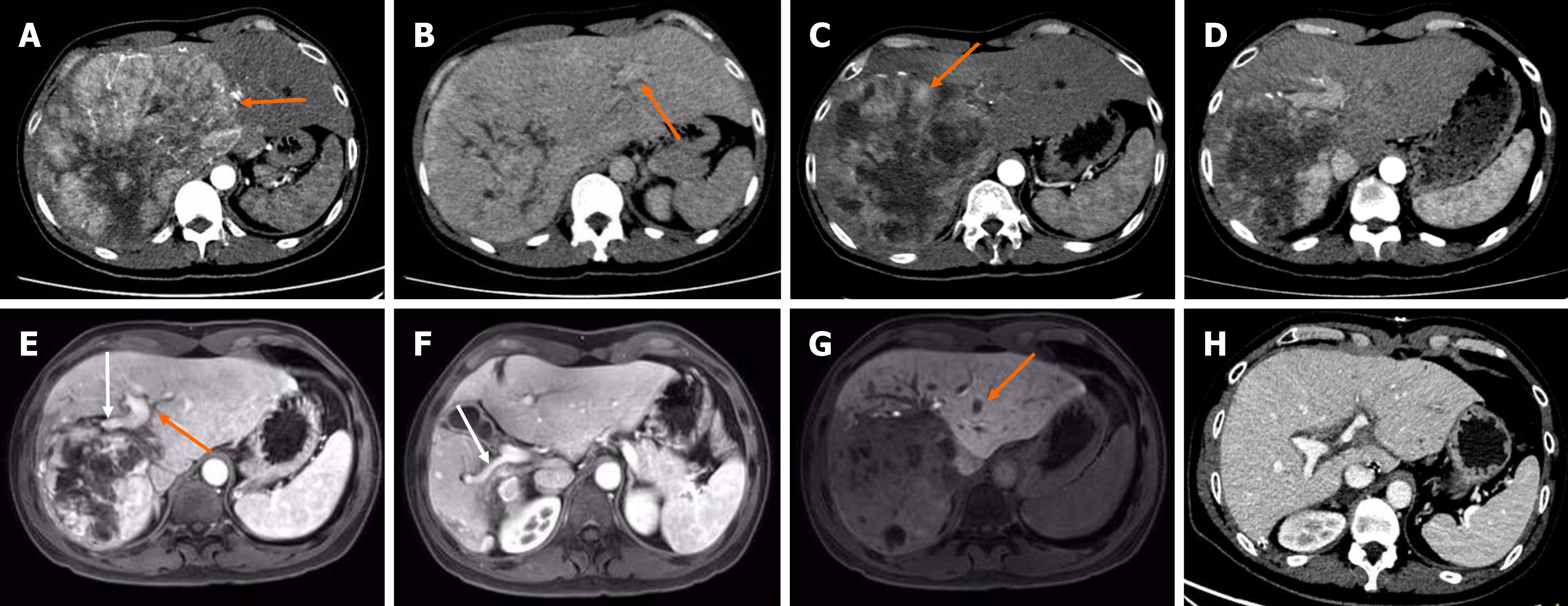
Figure 1 Imaging characteristics of the patient.
A: The arterial phase showed tumor had marked enhancement and abundant arterial blood supply (orange arrow); B: The portal venous phase showed the visualization of the left portal vein (orange arrow), but an absence of the right portal vein and the right hepatic vein, indicating a vascular invasion. Enhanced computed tomography (CT) showed hepatocellular carcinoma located in the right-left lobe of liver at initial diagnosis; C: After 4 cycles of drug-eluting bead transarterial chemoembolization plus hepatic arterial infusion chemotherapy, the tumor size was reduced but the residual tumor was still active (orange arrow); D: Four months after the first selective internal radiation therapy (SIRT), the residual liver volume increased progressively and the tumor volume significantly decreased; E-G: Two months after the second SIRT, gadoxetic acid-enhanced magnetic resonance imaging showed a further compensatory increase in left liver volume, a decrease in tumor volume, and the visualization of the left portal vein (E, orange arrow), the right anterior portal vein branch (E, white arrow) and the right posterior portal vein branch (F, white arrow), indicating the regression of the tumor thrombus and the recanalization of the portal vein flow. However, a new tumor was found in the left liver (G, orange arrow) at hepatobiliary phase; H: Five months after transplantation, the transplanted liver was well without tumor recurrence. A-D and H were enhanced CT scan.
- Citation: Liang LC, Huang WS, Guo ZX, You HJ, Guo YJ, Cai MY, Lin LT, Wang GY, Zhu KS. Liver transplantation following two conversions in a patient with huge hepatocellular carcinoma and portal vein invasion: A case report. World J Gastroenterol 2024; 30(36): 4071-4077
- URL: https://www.wjgnet.com/1007-9327/full/v30/i36/4071.htm
- DOI: https://dx.doi.org/10.3748/wjg.v30.i36.4071


