Copyright
©The Author(s) 2024.
World J Gastroenterol. Sep 28, 2024; 30(36): 4014-4020
Published online Sep 28, 2024. doi: 10.3748/wjg.v30.i36.4014
Published online Sep 28, 2024. doi: 10.3748/wjg.v30.i36.4014
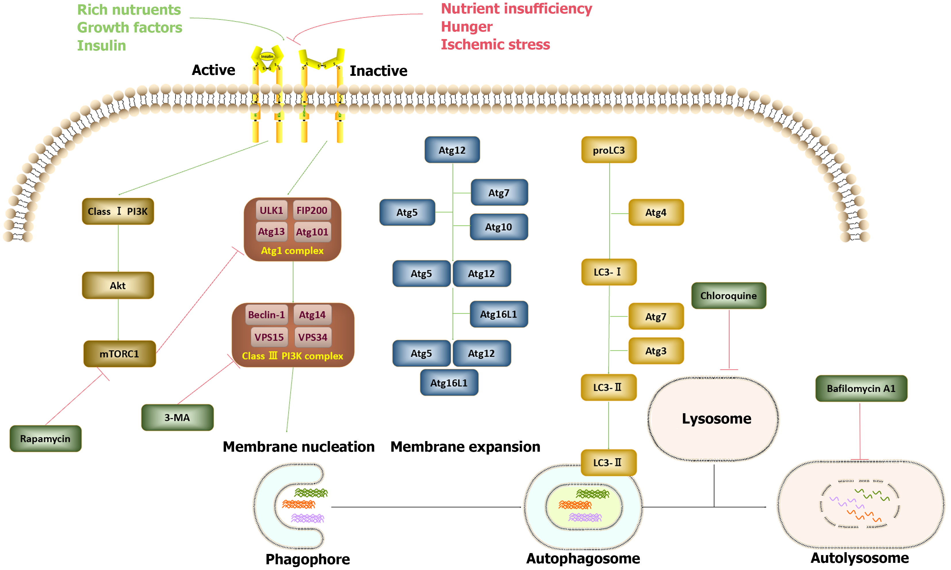
Figure 1 Illustrative diagram elucidating the signaling pathways governing the initiation and regulation of autophagy.
Autophagy, a cellular process, is stimulated under conditions such as nutrient deficiency, starvation, or ischemic stress. The initiation of autophagy involves distinct groups of autophagy-related gene (Atg) proteins. The formation of the Atg1 complex, comprising ULK1, focal adhesion kinase family kinase-interacting protein of 200 kDa, ATG13, and ATG101, triggers the assembly of the class III phosphatidylinositol 3-kinase (PI3K) complex, encompassing beclin-1, ATG14, VSP15, and VSP34, thereby initiating membrane nucleation and phagophore formation. Subsequent membrane expansion and fusion, facilitated by ATG5-ATG12-ATG16L1 and light chain 3-II, give rise to the autophagosome, which subsequently fuses with a lysosome, resulting in the establishment of a functional autophagy unit. Conversely, under conditions characterized by ample nutrients, growth factors, and insulin, activation of the class I PI3K-Akt- mechanistic target of rapamycin complex 1 (mTORC1) signaling pathway inhibits the formation of the Atg1 complex. While rapamycin serves as an autophagy inducer by inhibiting mTORC1, 3-MA, bafilomycin A1, and chloroquine obstruct the autophagy process through varied mechanisms. PI3K: Phosphatidylinositol 3-kinase.
- Citation: Shao BZ, Zhang WG, Liu ZY, Linghu EQ. Autophagy and its role in gastrointestinal diseases. World J Gastroenterol 2024; 30(36): 4014-4020
- URL: https://www.wjgnet.com/1007-9327/full/v30/i36/4014.htm
- DOI: https://dx.doi.org/10.3748/wjg.v30.i36.4014


