Copyright
©The Author(s) 2024.
World J Gastroenterol. Sep 21, 2024; 30(35): 3932-3941
Published online Sep 21, 2024. doi: 10.3748/wjg.v30.i35.3932
Published online Sep 21, 2024. doi: 10.3748/wjg.v30.i35.3932
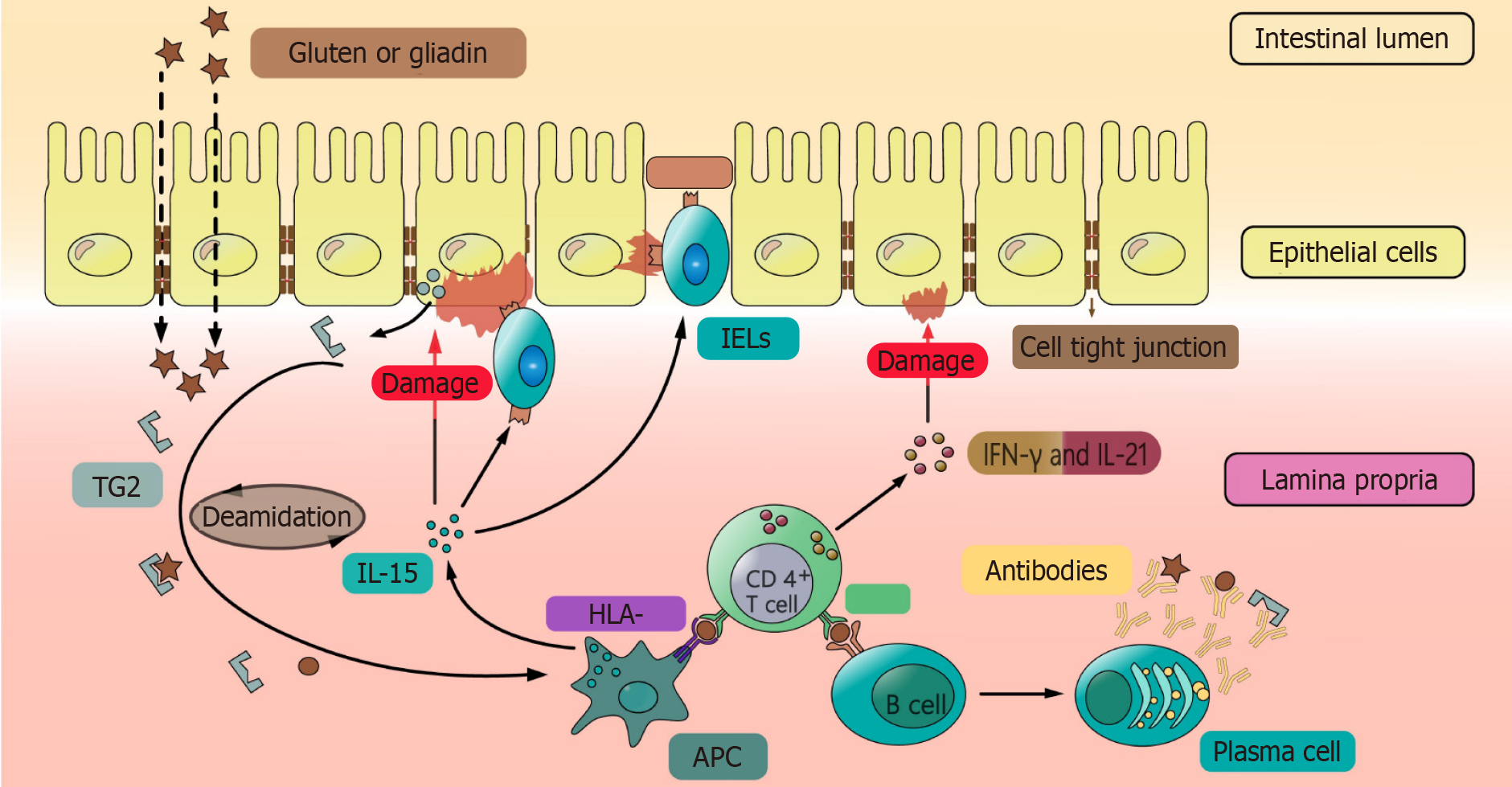
Figure 1 Pathophysiological mechanisms of celiac disease.
Gluten or gliadin crosses the epithelial cells, and after the deamidation by transglutaminase-2 (TG2), they become easier to be recognized by antigen presenting cells (APCs) expressing HLA-DQ2/8. APCs could secrete interleukin (IL)-15 which causes damage to epithelial cells directly or through activation of intraepithelial lymphocytes expressing natural killer cell receptor. Also, APCs present the deamidated polypeptides complex to CD4+ T cells which further secrete interferon-γ and IL-21 to cause damage to epithelial cells. In another way, CD4+ T cells activate B cells to differentiate into plasma cells with secretion of antibodies against gluten, TG2, and deamidated polypeptides complex. TG2: Transglutaminase-2; IL: Interleukin; HLA: Human leukocyte antigen; APC: Antigen presenting cells; TCR: T cell receptor; IELs: Intraepithelial lymphocytes; IFN-γ: Interferon-γ; NKG2D: Natural killer cell receptor.
- Citation: Ge HJ, Chen XL. Advances in understanding and managing celiac disease: Pathophysiology and treatment strategies. World J Gastroenterol 2024; 30(35): 3932-3941
- URL: https://www.wjgnet.com/1007-9327/full/v30/i35/3932.htm
- DOI: https://dx.doi.org/10.3748/wjg.v30.i35.3932


