Copyright
©The Author(s) 2024.
World J Gastroenterol. Aug 21, 2024; 30(31): 3717-3725
Published online Aug 21, 2024. doi: 10.3748/wjg.v30.i31.3717
Published online Aug 21, 2024. doi: 10.3748/wjg.v30.i31.3717
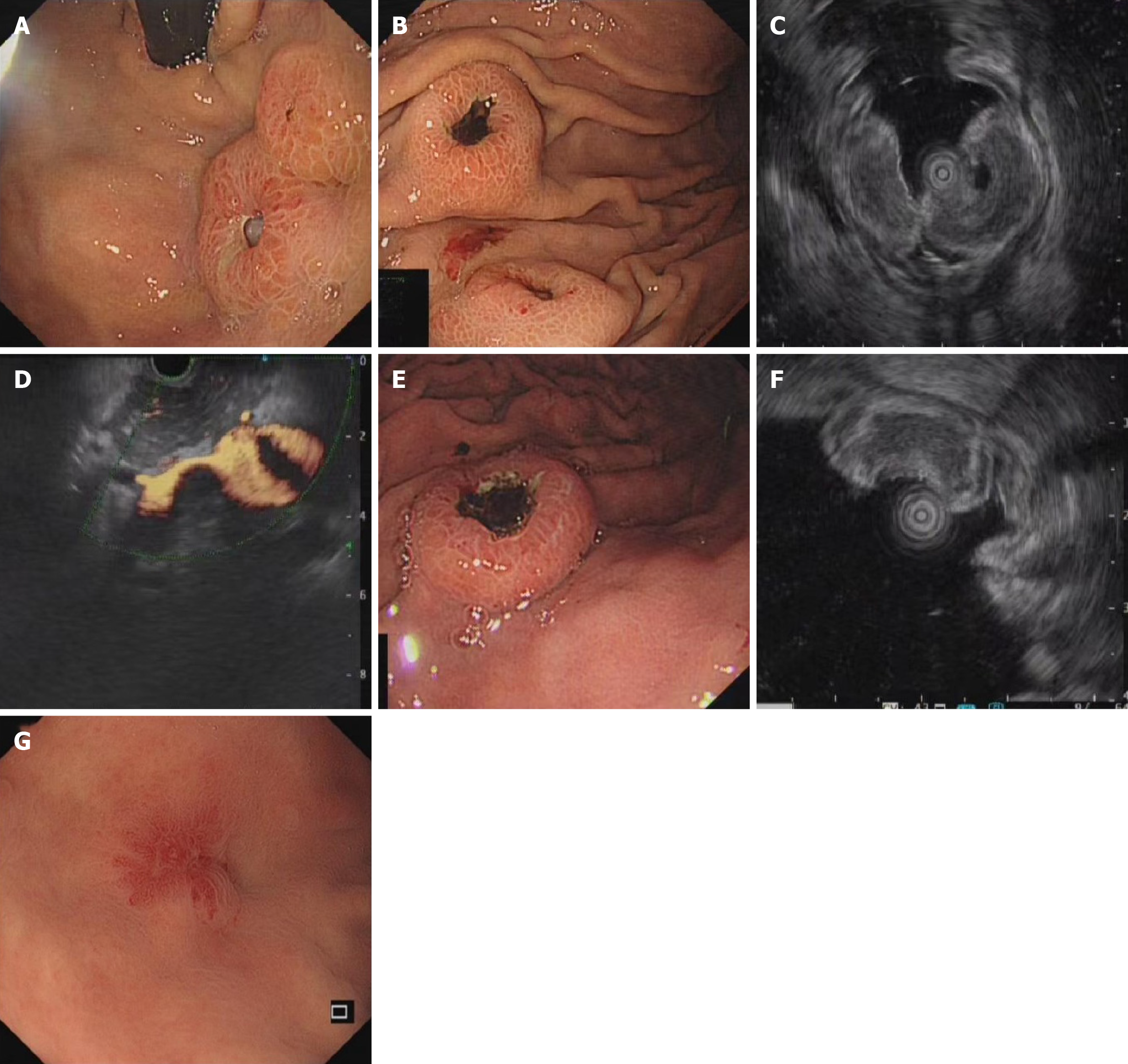
Figure 2 Gastroscopy and endoscopic ultrasound findings.
A and B: Gastroscopy suggested submucosal lesions with two central depressions and ulcer formation in the body and fundus of the stomach, respectively; C: Endoscopic ultrasound (EUS) suggested submucosal hypoechoic lesion of the gastric body; D: EUS suggested hypoechoic lesions of the portal vein (A-D; case 1); E: Gastroscopy suggested a submucosal lesion with ulcer formation was seen in the greater curvature of the stomach; F: EUS showed a submucosal hypoechoic lesion in the greater curvature of the stomach (E and F; case 2); G: A 4 mm × 5 mm mucosal lesion with a depressed tip (G; case 3).
- Citation: Yang S, He QY, Zhao QJ, Yang HT, Yang ZY, Che WY, Li HM, Wu HC. Gastric metastasis of small cell lung carcinoma: Three case reports and review of literature. World J Gastroenterol 2024; 30(31): 3717-3725
- URL: https://www.wjgnet.com/1007-9327/full/v30/i31/3717.htm
- DOI: https://dx.doi.org/10.3748/wjg.v30.i31.3717


