Copyright
©The Author(s) 2024.
World J Gastroenterol. Aug 21, 2024; 30(31): 3689-3704
Published online Aug 21, 2024. doi: 10.3748/wjg.v30.i31.3689
Published online Aug 21, 2024. doi: 10.3748/wjg.v30.i31.3689
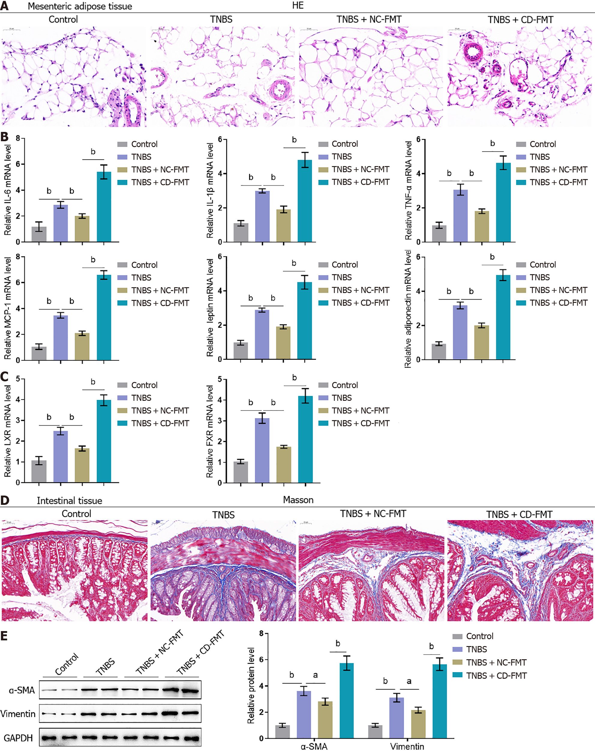
Figure 5 Effects of gut microbiota from Crohn’s disease patients on the histopathological characteristics of mesenteric adipose and intestinal tissues from mice with 2,4,6-trinitrobenzene sulfonic acid-induced Crohn’s disease.
A: Mesenteric adipose tissues collected from mice were stained with hematoxylin-eosin to assess histopathological alterations (× 150); B and C: Real-time reverse transcriptase-polymerase chain reaction assays of interleukin-6 (IL-6), IL-1β, tumour necrosis factor alpha, MCP-1, leptin, adiponectin, LXR, and FXR mRNA levels in mouse tissues; D: Intestinal tissues from mice stained with Masson stain to evaluate fibrotic alterations (× 150); E: Immunoblotting assays of alpha-smooth muscle actin and vimentin protein levels in mouse intestinal tissues. n = 6 or 3, bP < 0.01 vs control; aP < 0.05, bP < 0.01 vs 2,4,6-trinitrobenzene sulfonic acid (TNBS) alone; bP < 0.01 vs TNBS + NC-fetal microbiota transplantation, by one-way ANOVA with post-hoc Tukey HSD test. TNBS: 2,4,6-trinitrobenzene sulfonic acid; CD: Crohn’s disease; NC: Normal control; FMT: Fetal microbiota transplantation; IFN-γ: Interferon-gamma; TNF-α: Tumour necrosis factor alpha; IL: Interleukin; HE: Hematoxylin and eosin; α-SMA: Alpha-smooth muscle actin.
- Citation: Wu Q, Yuan LW, Yang LC, Zhang YW, Yao HC, Peng LX, Yao BJ, Jiang ZX. Role of gut microbiota in Crohn’s disease pathogenesis: Insights from fecal microbiota transplantation in mouse model. World J Gastroenterol 2024; 30(31): 3689-3704
- URL: https://www.wjgnet.com/1007-9327/full/v30/i31/3689.htm
- DOI: https://dx.doi.org/10.3748/wjg.v30.i31.3689


