Copyright
©The Author(s) 2024.
World J Gastroenterol. Jun 28, 2024; 30(24): 3086-3105
Published online Jun 28, 2024. doi: 10.3748/wjg.v30.i24.3086
Published online Jun 28, 2024. doi: 10.3748/wjg.v30.i24.3086
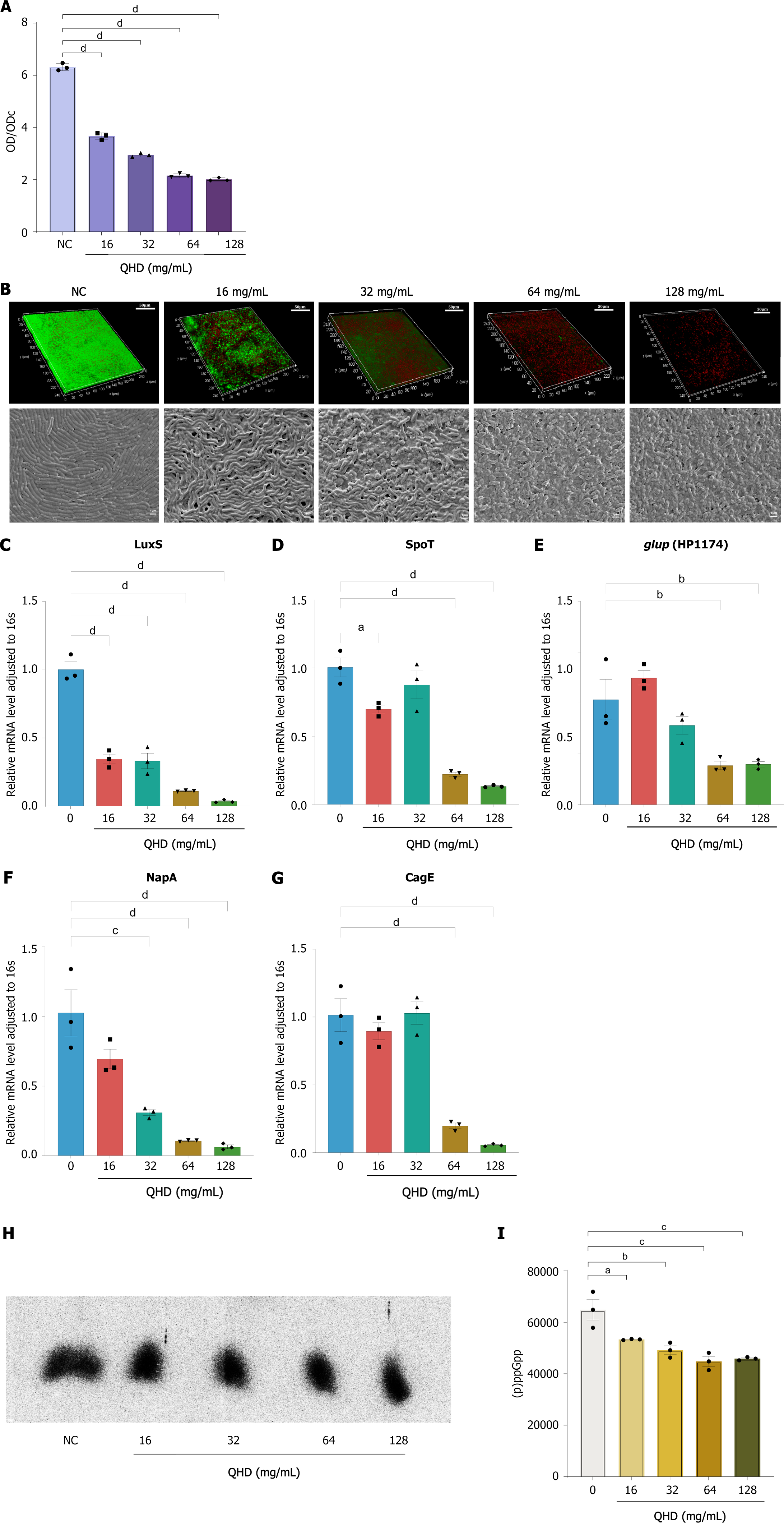
Figure 5 Qingre Huashi decoction inhibits and disrupts Helicobacter pylori biofilm formation in vitro.
A: Crystal violet staining indicated that Qingre Huashi decoction (QHD) inhibited the in vitro biofilm formation of Helicobacter pylori (HP) (16-128 mg/mL) in a concentration-dependent manner, with significant inhibition observed at a concentration of 16 mg/mL (1/4 MIC); B: CLSM analysis revealed that Strain 1 exhibited strong viability and significant bacterial activity in vitro, causing the formation of a dense biofilm. Treatment with various concentrations (16-128 mg/mL) of QHD resulted in a significant decrease in the viability of HP, reduced activity, and the formation of biofilms that were thinner and less tightly structured. Based on scanning electron microscopy, the NC group exemplified the typical biofilm arrangement of HP in a laboratory setting, characterized by predominantly rod-shaped bacteria that were densely and closely packed, which could form a cohesive biofilm matrix. After the administration of QHD, the attachment of bacteria to the biofilm was weakened, and the structure of the biofilm became fragmented, displaying multiple empty spaces; C-G: Expression levels of biofilm-related genes LuxS, NapA, Spot, HP1174, and CagE examined by quantitative real-time polymerase chain reaction before and after QHD intervention. The results found that QHD could inhibit the expression of these biofilm-related genes, thereby affecting the formation of biofilms; H and I: 32P-labeled nucleotides in HP detected by thin-layer chromatography after drug intervention. QHD significantly inhibited HP (p)ppGpp molecule expression. QHD: Qingre Huashi decoction; OD: Optical density; SEM: Scanning electron microscopy. aP < 0.05; bP < 0.01; cP < 0.001; dP < 0.0001.
- Citation: Lin MM, Yang SS, Huang QY, Cui GH, Jia XF, Yang Y, Shi ZM, Ye H, Zhang XZ. Effect and mechanism of Qingre Huashi decoction on drug-resistant Helicobacter pylori. World J Gastroenterol 2024; 30(24): 3086-3105
- URL: https://www.wjgnet.com/1007-9327/full/v30/i24/3086.htm
- DOI: https://dx.doi.org/10.3748/wjg.v30.i24.3086


