Copyright
©The Author(s) 2024.
World J Gastroenterol. Jun 21, 2024; 30(23): 3005-3015
Published online Jun 21, 2024. doi: 10.3748/wjg.v30.i23.3005
Published online Jun 21, 2024. doi: 10.3748/wjg.v30.i23.3005
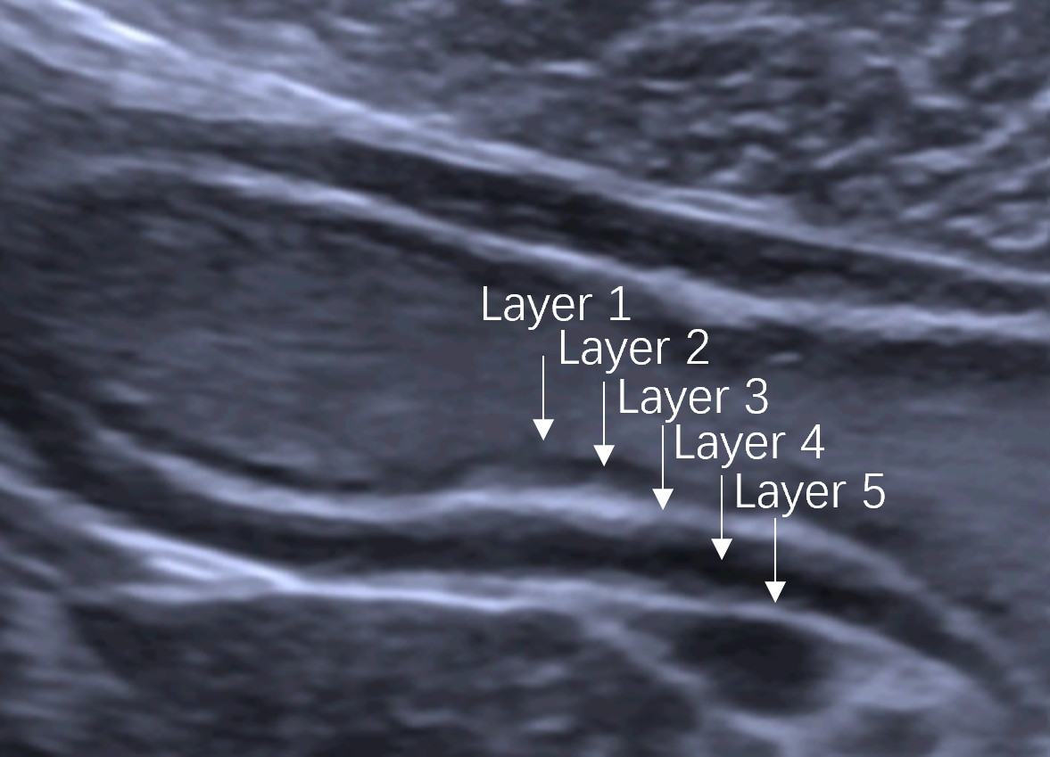
Figure 2 The 5-layer structure of the normal gastric wall on ultrasonography.
The 5-layer structure of the normal gastric wall is numbered from the luminal side. Layer 1 is the interface echo between the gastric lumen and the mucosa, layer 2 is the rest of the mucosa, layer 3 is the submucosa, layer 4 is the muscularis propria, and layer 5 is the serosa.
- Citation: Xu YF, Ma HY, Huang GL, Zhang YT, Wang XY, Wei MJ, Pei XQ. Double contrast-enhanced ultrasonography improves diagnostic accuracy of T staging compared with multi-detector computed tomography in gastric cancer patients. World J Gastroenterol 2024; 30(23): 3005-3015
- URL: https://www.wjgnet.com/1007-9327/full/v30/i23/3005.htm
- DOI: https://dx.doi.org/10.3748/wjg.v30.i23.3005


