Copyright
©The Author(s) 2024.
World J Gastroenterol. Jan 14, 2024; 30(2): 137-145
Published online Jan 14, 2024. doi: 10.3748/wjg.v30.i2.137
Published online Jan 14, 2024. doi: 10.3748/wjg.v30.i2.137
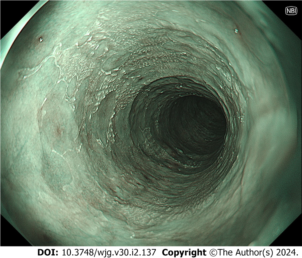
Figure 3 Narrow band imaging shows “epidermization”, which was widely spread in the middle esophagus 2.
5 years after the first endoscopic examination. The extent of epidermization has not markedly changed over a 3-year period. Olympus GIF-XZ1200.
- Citation: Shintaku M. Esophageal intramural pseudodiverticulosis. World J Gastroenterol 2024; 30(2): 137-145
- URL: https://www.wjgnet.com/1007-9327/full/v30/i2/137.htm
- DOI: https://dx.doi.org/10.3748/wjg.v30.i2.137


