Copyright
©The Author(s) 2024.
World J Gastroenterol. Jan 14, 2024; 30(2): 137-145
Published online Jan 14, 2024. doi: 10.3748/wjg.v30.i2.137
Published online Jan 14, 2024. doi: 10.3748/wjg.v30.i2.137
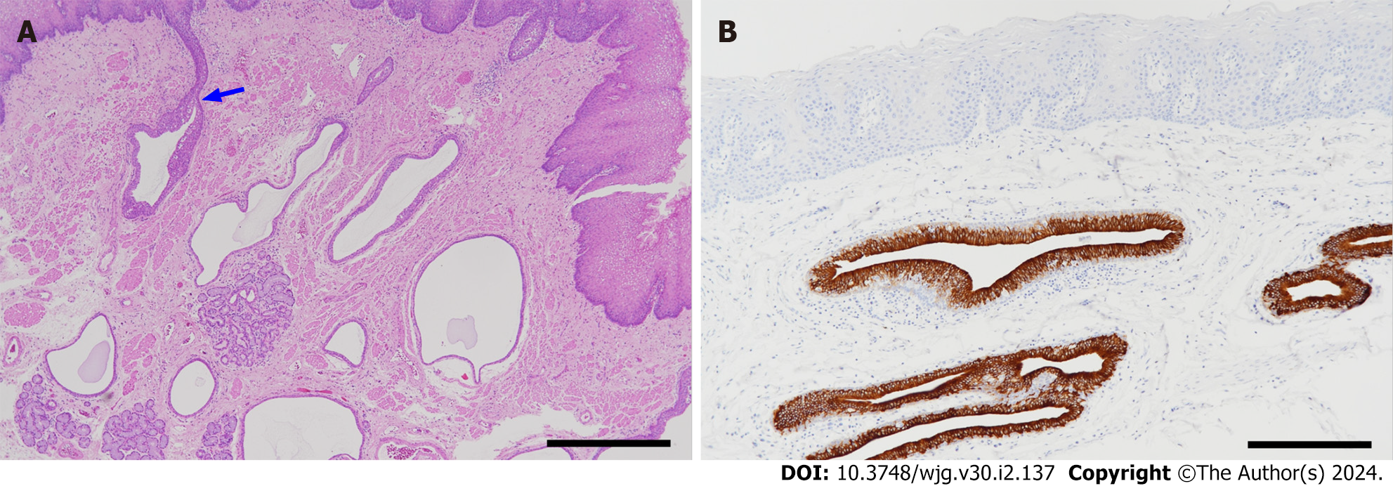
Figure 2 Pathological findings.
A: Histology of esophageal intramural pseudodiverticulosis (hematoxylin-eosin stain). Many cystically dilated ducts of the esophageal glands are seen in the lamina propria mucosae and submucosa. Some of them are continuous with the superficial epithelium (arrow) (scale bar: 1 mm); B: Cytokeratin 7 (CK7) immunostaining. Epithelial cells lining the ducts are positive for CK7. The epithelium of the duct shows hyperplasia and stratification. The esophageal mucosal epithelium (upper part of the figure) is negative for CK7 (scale bar: 200 µm).
- Citation: Shintaku M. Esophageal intramural pseudodiverticulosis. World J Gastroenterol 2024; 30(2): 137-145
- URL: https://www.wjgnet.com/1007-9327/full/v30/i2/137.htm
- DOI: https://dx.doi.org/10.3748/wjg.v30.i2.137


