Copyright
©The Author(s) 2024.
World J Gastroenterol. Jan 14, 2024; 30(2): 137-145
Published online Jan 14, 2024. doi: 10.3748/wjg.v30.i2.137
Published online Jan 14, 2024. doi: 10.3748/wjg.v30.i2.137
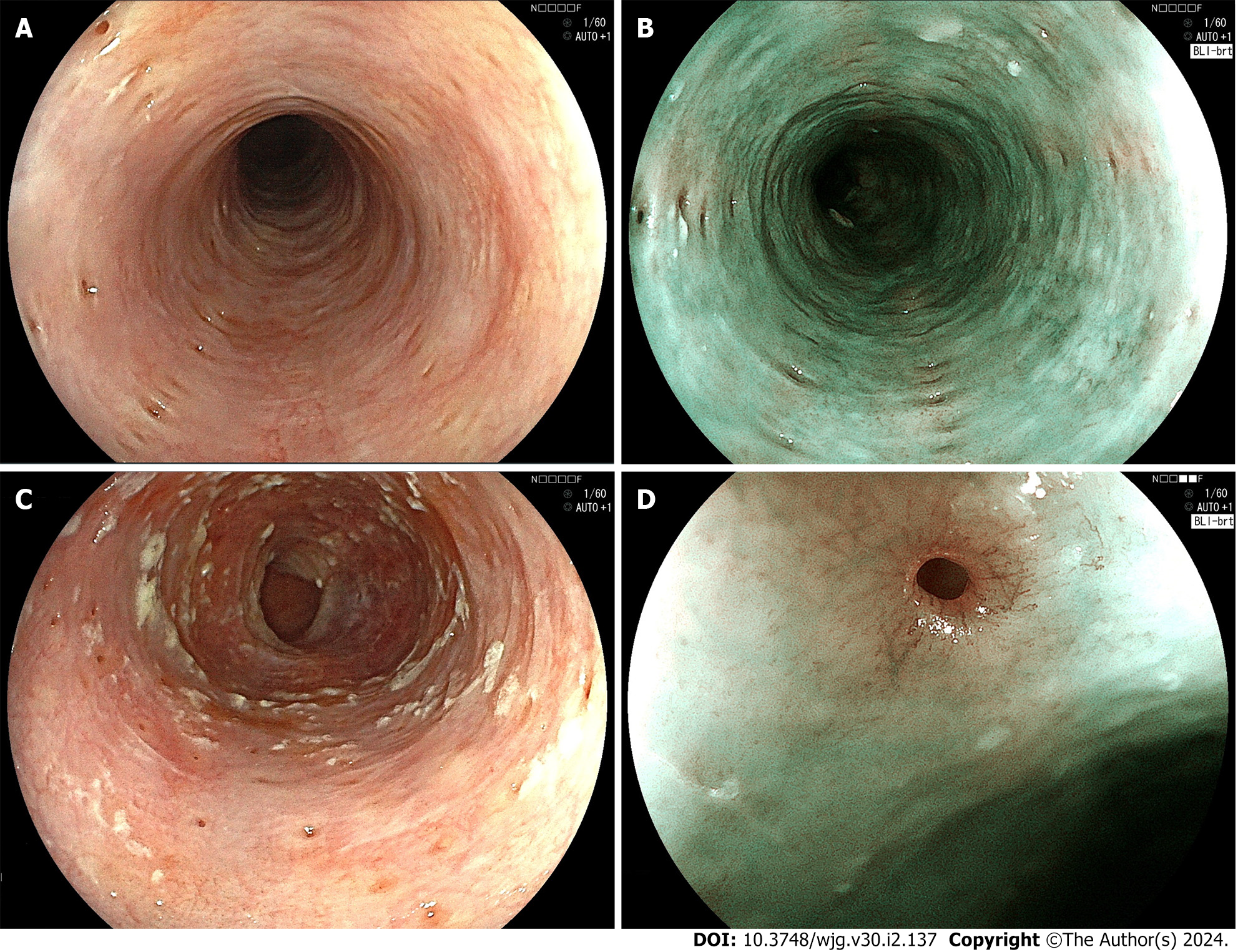
Figure 1 Endoscopic examination.
A: White-light endoscopic image. Multiple small diverticulum-like depressions of the mucosa with a diameter of 1-4 mm are seen. Fujifilm EG-L600ZW; B: Blue laser imaging (BLI)-bright image. The light-brown depressions are longitudinally aligned on the green-colored mucosa. Fujifilm EG-L600ZW; C: White-light endoscopic image. The orifices of pseudodiverticula are covered with whitish, plaque-like material. Fujifilm EG-L600ZW; D: BLI-bright, medium magnifying image. The orifice of the pseudodiverticulum is surrounded by blood vessels arranged in an eyelash-like pattern. Fujifilm EG-L600ZW.
- Citation: Shintaku M. Esophageal intramural pseudodiverticulosis. World J Gastroenterol 2024; 30(2): 137-145
- URL: https://www.wjgnet.com/1007-9327/full/v30/i2/137.htm
- DOI: https://dx.doi.org/10.3748/wjg.v30.i2.137


