Copyright
©The Author(s) 2024.
World J Gastroenterol. May 21, 2024; 30(19): 2523-2537
Published online May 21, 2024. doi: 10.3748/wjg.v30.i19.2523
Published online May 21, 2024. doi: 10.3748/wjg.v30.i19.2523
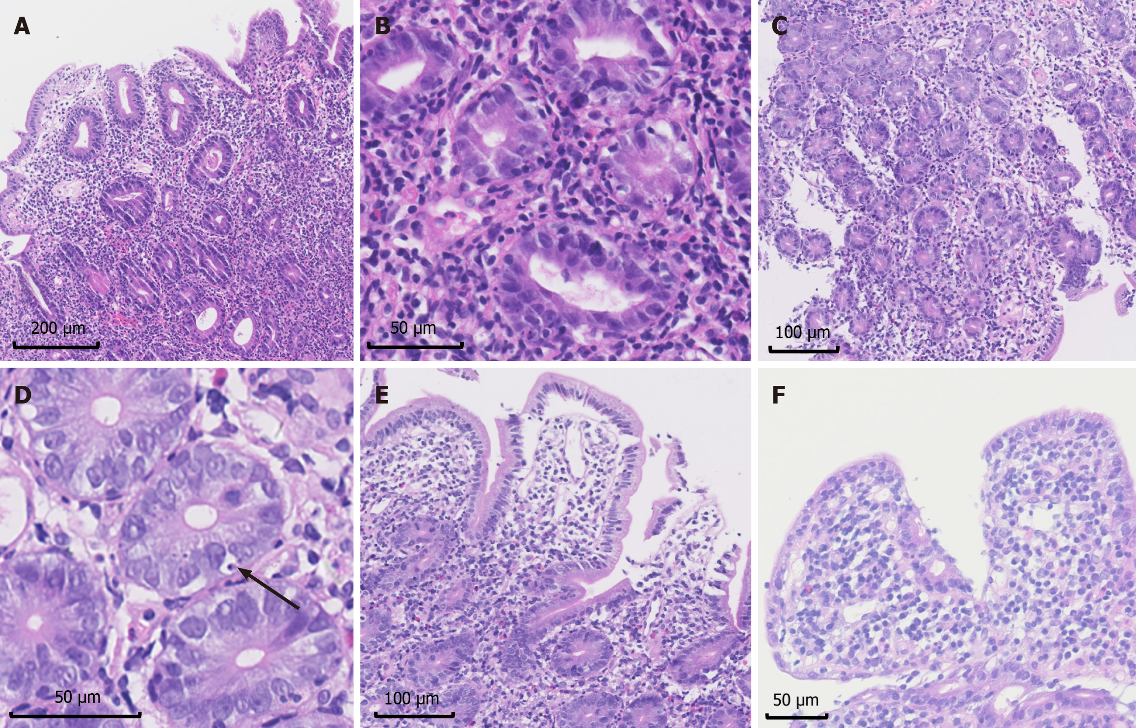
Figure 6 Images of characteristic histological manifestations in autoimmune enteropathy patients (hematoxylin and eosin staining).
A: Villous blunting (40 ×); B: Mononuclear inflammatory infiltration (200 ×); C: Absent goblet cells (40 ×); D: Increased apoptotic bodies (black arrow, 400 ×); E: Minimal intraepithelial lymphocytosis (200 ×); F: Intraepithelial lymphocytosis (200 ×).
- Citation: Li MH, Ruan GC, Zhou WX, Li XQ, Zhang SY, Chen Y, Bai XY, Yang H, Zhang YJ, Zhao PY, Li J, Li JN. Clinical manifestations, diagnosis and long-term prognosis of adult autoimmune enteropathy: Experience from Peking Union Medical College Hospital. World J Gastroenterol 2024; 30(19): 2523-2537
- URL: https://www.wjgnet.com/1007-9327/full/v30/i19/2523.htm
- DOI: https://dx.doi.org/10.3748/wjg.v30.i19.2523


