Copyright
©The Author(s) 2024.
World J Gastroenterol. May 14, 2024; 30(18): 2418-2439
Published online May 14, 2024. doi: 10.3748/wjg.v30.i18.2418
Published online May 14, 2024. doi: 10.3748/wjg.v30.i18.2418
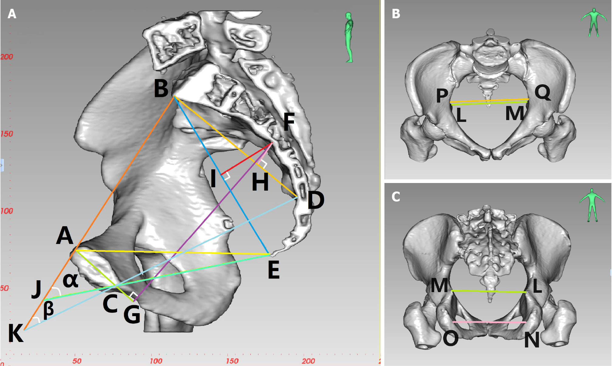
Figure 2 Diagram of three-dimensional reconstruction of the pelvis in a female patient.
A: Midsagittal lateral view: anterior-posterior diameter of pelvic inlet (AB), anterior-posterior diameter of mid-pelvis (CD), anterior-posterior diameter of pelvic outlet (CE), superior-inferior diameter of the pubic symphysis (AC), sacrococcygeal distance (BE), superior-inferior diameter of sacrum (BD), superior pubococcygeal diameter (AE), anterior-posterior sacropubic distance (FG), sacrococcygeal-pubic angle (α), and sacropubic angle (β); B: Anterior-posterior view: transverse diameter of pelvic inlet (PQ), interischial spine diameter (LM); C: Posterior-anterior view: interischial spine diameter (LM), interischial tuberosity diameter (NO).
- Citation: Zhou XC, Guan SW, Ke FY, Dhamija G, Wang Q, Chen BF. Construction of a nomogram model to predict technical difficulty in performing laparoscopic sphincter-preserving radical resection for rectal cancer. World J Gastroenterol 2024; 30(18): 2418-2439
- URL: https://www.wjgnet.com/1007-9327/full/v30/i18/2418.htm
- DOI: https://dx.doi.org/10.3748/wjg.v30.i18.2418


