Copyright
©The Author(s) 2024.
World J Gastroenterol. Apr 28, 2024; 30(16): 2195-2208
Published online Apr 28, 2024. doi: 10.3748/wjg.v30.i16.2195
Published online Apr 28, 2024. doi: 10.3748/wjg.v30.i16.2195
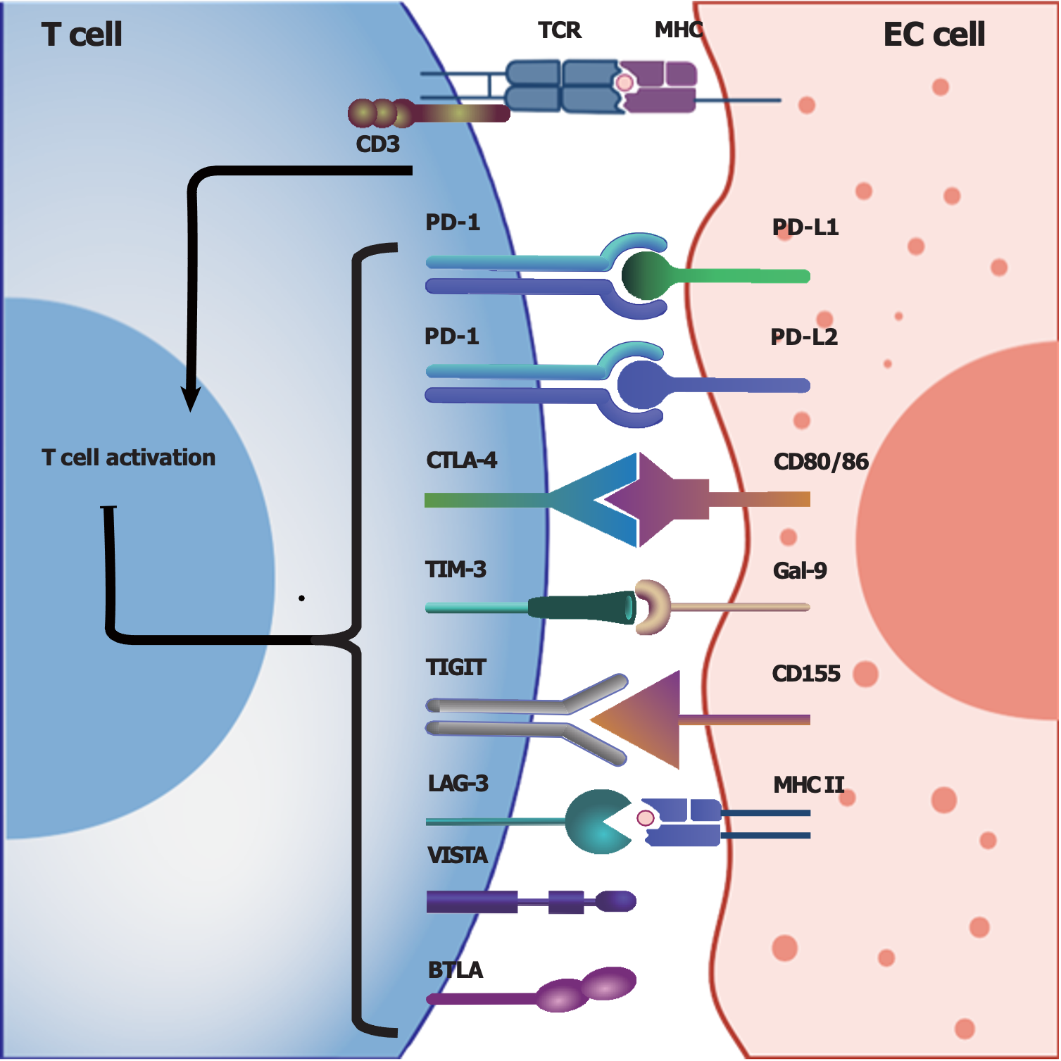
Figure 1 Summary of potentially involved immune checkpoints in esophageal cancer.
T cells can be activated by interacting with major histocompatibility complexes expressed on esophageal cancer (EC) cells, and the presence and interaction of immune checkpoints with their ligands can suppress T-cell activation and function to achieve immunosuppression. Herein, we summarize the immune checkpoints and their ligands that are potentially involved in the tumor microenvironment of EC. Programmed cell death protein 1, cytolytic T lymphocyte-associated antigen-4, T-cell immunoglobulin and mucin-domain containing-3 (TIM-3), T-cell immunoglobulin and immunoreceptor tyrosine-based inhibitory motif domain (TIGIT), lymphocyte activation gene 3 (LAG-3), V-domain Ig suppressor of T-cell activation and B- and T-lymphocyte attenuator are expressed on T cells, while TIM-3, TIGIT and LAG-3 are also expressed on natural killer cells. EC: Esophageal cancer; TCR: T cell receptor; MHC: Major histocompatibility complex; CD: Cluster of differentiation; PD-1: Programmed cell death protein 1; PD-L1: Programmed cell death ligand 1; PD-L2: Programmed cell death ligand 2; CTLA-4: Cytolytic T lymphocyte-associated antigen-4; TIM-3: T-cell immunoglobulin and mucin-domain containing-3; Gal-9: Galectin-9; TIGIT: T-cell immunoglobulin and immunoreceptor tyrosine-based inhibitory motif domain; LAG-3: Lymphocyte activation gene 3; VISTA: V-domain Ig suppressor of T-cell activation; BTLA: B- and T-lymphocyte attenuator.
- Citation: Zhang XJ, Yu Y, Zhao HP, Guo L, Dai K, Lv J. Mechanisms of tumor immunosuppressive microenvironment formation in esophageal cancer. World J Gastroenterol 2024; 30(16): 2195-2208
- URL: https://www.wjgnet.com/1007-9327/full/v30/i16/2195.htm
- DOI: https://dx.doi.org/10.3748/wjg.v30.i16.2195


