Copyright
©The Author(s) 2024.
World J Gastroenterol. Apr 21, 2024; 30(15): 2155-2174
Published online Apr 21, 2024. doi: 10.3748/wjg.v30.i15.2155
Published online Apr 21, 2024. doi: 10.3748/wjg.v30.i15.2155
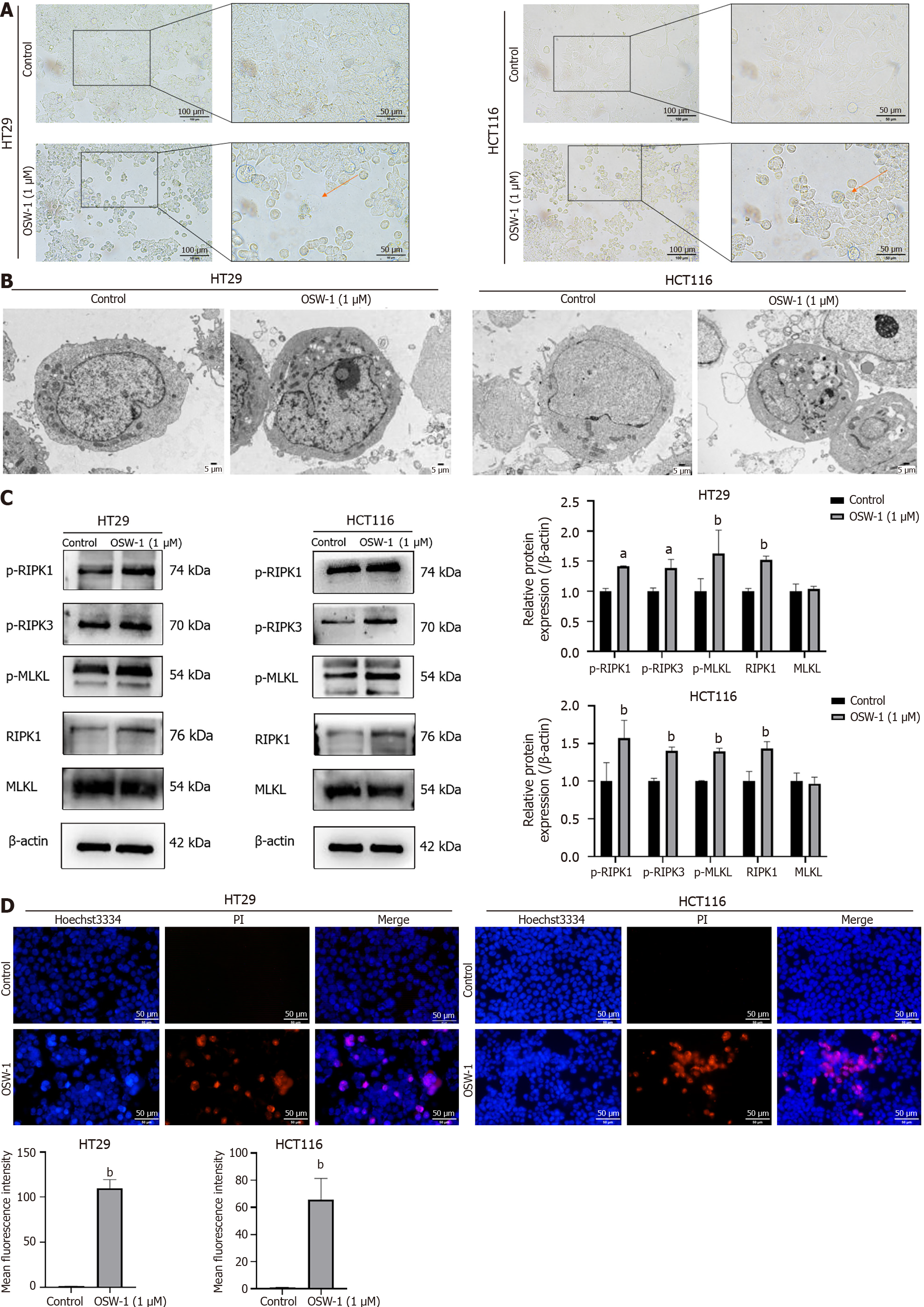
Figure 3 OSW-1 triggers necroptosis in colorectal cancer cell culture.
A: Necrotic morphological changes were observed in HT29 and HCT116 cells treated with OSW-1 using optical microscopy (400 × magnification). Necrotic cells are indicated by red arrows; B: Typical morphological changes associated with necroptosis were identified in HT29 and HCT116 cells treated with OSW-1 through TEM (3000 × magnification); C: The expression levels of proteins associated with necroptosis in HT29 and HCT116 cells following 24 h of exposure to OSW-1; D: A Hoechst 33342/propidium iodide dual staining assay was used to assess the rate of necroptosis in HT29 and HCT116 cells following treatment with OSW-1. Scale bar = 50 μm. aP < 0.05 and bP < 0.01 vs Control. Each data point represents the mean ± SE. PI: Propidium iodide.
- Citation: Wang N, Li CY, Yao TF, Kang XD, Guo HS. OSW-1 triggers necroptosis in colorectal cancer cells through the RIPK1/RIPK3/MLKL signaling pathway facilitated by the RIPK1-p62/SQSTM1 complex. World J Gastroenterol 2024; 30(15): 2155-2174
- URL: https://www.wjgnet.com/1007-9327/full/v30/i15/2155.htm
- DOI: https://dx.doi.org/10.3748/wjg.v30.i15.2155


