Copyright
©The Author(s) 2024.
World J Gastroenterol. Apr 14, 2024; 30(14): 2059-2067
Published online Apr 14, 2024. doi: 10.3748/wjg.v30.i14.2059
Published online Apr 14, 2024. doi: 10.3748/wjg.v30.i14.2059
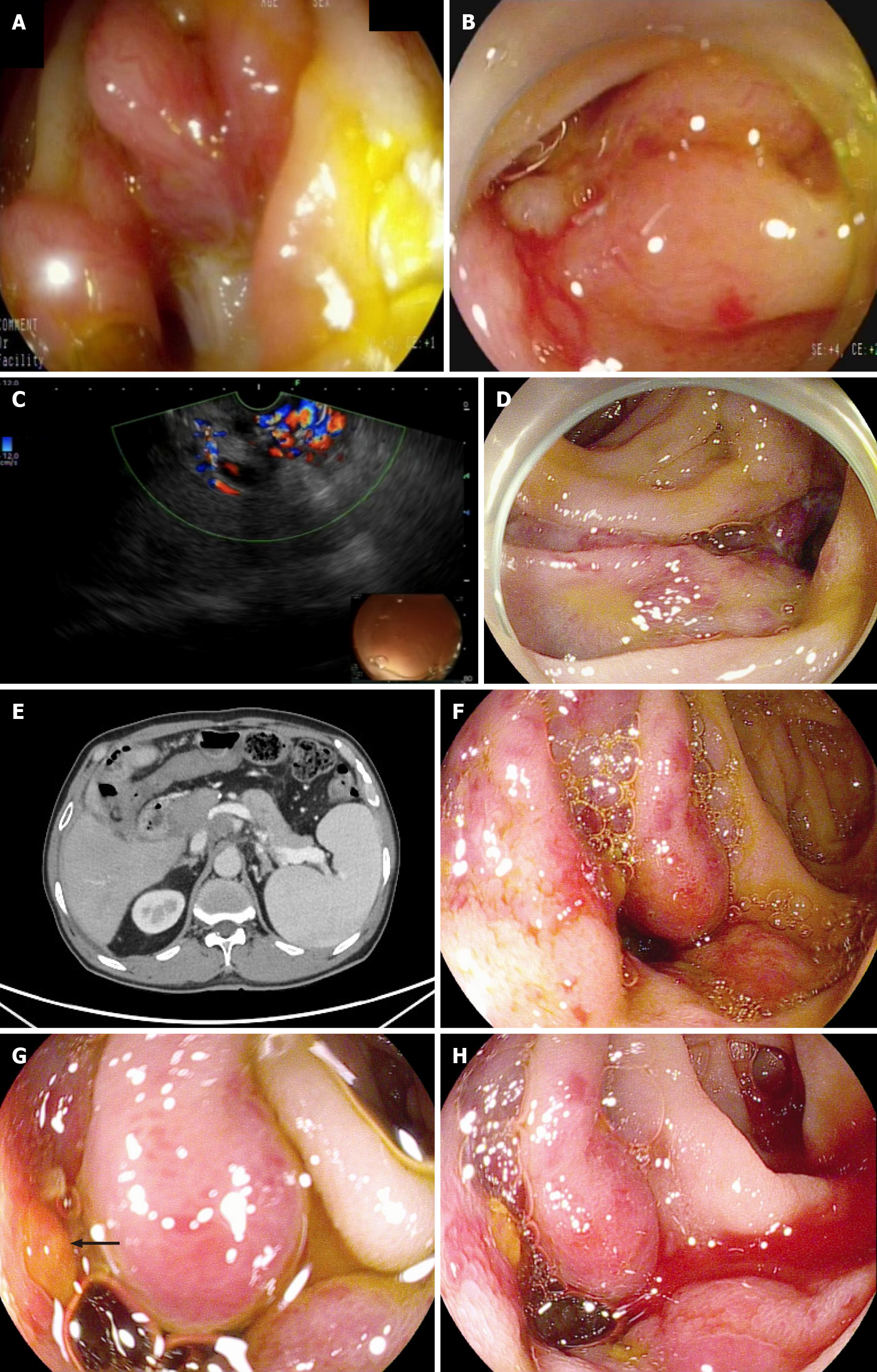
Figure 1 Emergency endoscopy confirmed the presence of numerous varicose veins before endoscopic sclerotherapy and was validated by endoscopic ultrasonography.
A: Emergency gastroscopy revealed tortuous dilated blood vessels around the choledochojejunostomy site (Case 1); B: A mucosal rupture was detected with spontaneous bleeding (Case 1); C: Endoscopic ultrasonography confirmed the presence of numerous varicose veins with abundant blood flow (Case 1); D: Emergency gastroscopy revealed a significant number of tortuous dilated varicose veins near the choledochojejunostomy site, along with several active bleedings. Hemostatic clips were used before endoscopic sclerotherapy (Case 2); E: Computed tomography showed prehepatic portal hypertension, suspicious tumor recurrence in the hepatic hilum area and the formation of varices around the choledochojejunostomy site (Case 3); F: Colonoscopy revealed three varicose veins visible with signs of erythema (Case 3); G: Thrombus head (arrow) was detected (Case 3); H: Spontaneous bleeding was observed at the site of varicose veins (Case 3).
- Citation: Liu J, Wang P, Wang LM, Guo J, Zhong N. Outcomes of endoscopic sclerotherapy for jejunal varices at the site of choledochojejunostomy (with video): Three case reports. World J Gastroenterol 2024; 30(14): 2059-2067
- URL: https://www.wjgnet.com/1007-9327/full/v30/i14/2059.htm
- DOI: https://dx.doi.org/10.3748/wjg.v30.i14.2059


