Copyright
©The Author(s) 2024.
World J Gastroenterol. Mar 14, 2024; 30(10): 1431-1449
Published online Mar 14, 2024. doi: 10.3748/wjg.v30.i10.1431
Published online Mar 14, 2024. doi: 10.3748/wjg.v30.i10.1431
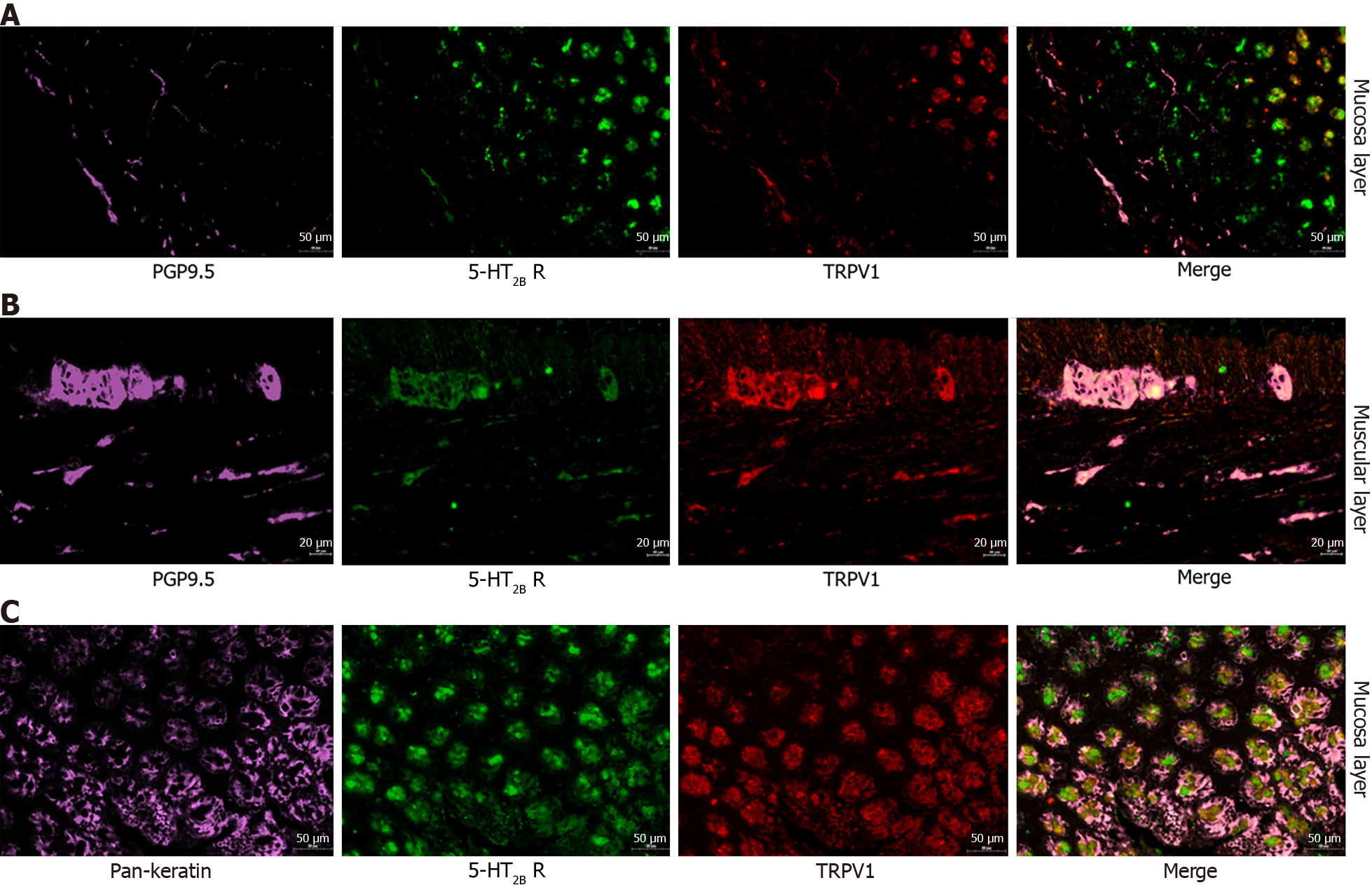
Figure 7 Immunofluorescence analysis of colon tissues costained with serotonin receptor 2B (green fluorescence), transient receptor potential vanilloid type 1 (red fluorescence), protein gene product 9.
5 (purple fluorescence) and Pan-Keratin (purple fluorescence). A: Serotonin receptor 2B (5-HT2B receptor) receptor (green fluorescence), transient receptor potential vanilloid type 1 (TRPV1) (red fluorescence) and protein gene product 9.5 (PGP9.5) (purple fluorescence) in the rat colon mucosa layer. Merged image showing colocalization (pink) of the 5-HT2B receptor, TRPV1 receptor and PGP9.5 immunoreactivities in the rat colon mucosa layer; B: 5-HT2B receptor (green fluorescence), TRPV1 (red fluorescence) and PGP9.5 (purple fluorescence) in the rat colon muscular layer. Merged image showing colocalization (pink) of the 5-HT2B receptor, TRPV1 receptor and PGP9.5 immunoreactivities in the rat colon muscular layer; C: The 5-HT2B receptor (green fluorescence), TRPV1 (red fluorescence) and Pan-Keratin (purple fluorescence) in the rat colon mucosa layer. Merged image showing colocalization (pink) of the 5-HT2B receptor and TRPV1 and pankeratin immunoreactivities in the rat colon mucosa layer. Magnification 20 ×. PGP9.5: Protein gene product 9.5; 5-HT2B: Serotonin receptor 2B; TRPV1: Transient receptor potential vanilloid type 1.
- Citation: Li ZY, Mao YQ, Hua Q, Sun YH, Wang HY, Ye XG, Hu JX, Wang YJ, Jiang M. Serotonin receptor 2B induces visceral hyperalgesia in rat model and patients with diarrhea-predominant irritable bowel syndrome. World J Gastroenterol 2024; 30(10): 1431-1449
- URL: https://www.wjgnet.com/1007-9327/full/v30/i10/1431.htm
- DOI: https://dx.doi.org/10.3748/wjg.v30.i10.1431


