Copyright
©The Author(s) 2024.
World J Gastroenterol. Mar 14, 2024; 30(10): 1405-1419
Published online Mar 14, 2024. doi: 10.3748/wjg.v30.i10.1405
Published online Mar 14, 2024. doi: 10.3748/wjg.v30.i10.1405
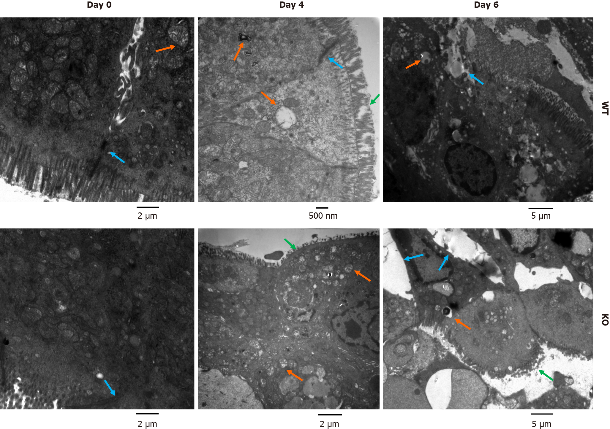
Figure 4 Ultrastructure of colonic mucosal epithelial cells in mice after dextran sulfate sodium induction.
Mice were treated with 3% dextran sulfate sodium (DSS) water. Colon sections (0.5 cm) were removed on days 0, 4, and 6 after DSS treatment and fixed in 2% glutaraldehyde. The microvilli, cell structure, organelles and cell junctions of intestinal mucosal epithelial cells were examined by electron microscopy. Before DSS induction, the junctions between epithelial cells in wild-type (WT) and gene knockout (KO) mice were normal. After DSS induction, WT mice exhibited a loss of microvilli, pyknotic nuclei and impaired tight junctions. In KO mice, severe colonic epithelial damage was observed, and the intestinal epithelial cells exhibited damaged microvilli, widening of intercellular spaces, nuclear pyknosis, and mitochondrial swelling and degeneration (green arrows show damaged microvilli, orange arrows show mitochondrial swelling, and blue arrows show the intercellular space).
- Citation: Tian Y, Li X, Wang X, Pei ST, Pan HX, Cheng YQ, Li YC, Cao WT, Petersen JDD, Zhang P. Alkaline sphingomyelinase deficiency impairs intestinal mucosal barrier integrity and reduces antioxidant capacity in dextran sulfate sodium-induced colitis. World J Gastroenterol 2024; 30(10): 1405-1419
- URL: https://www.wjgnet.com/1007-9327/full/v30/i10/1405.htm
- DOI: https://dx.doi.org/10.3748/wjg.v30.i10.1405


