Copyright
©The Author(s) 2024.
World J Gastroenterol. Jan 7, 2024; 30(1): 34-49
Published online Jan 7, 2024. doi: 10.3748/wjg.v30.i1.34
Published online Jan 7, 2024. doi: 10.3748/wjg.v30.i1.34
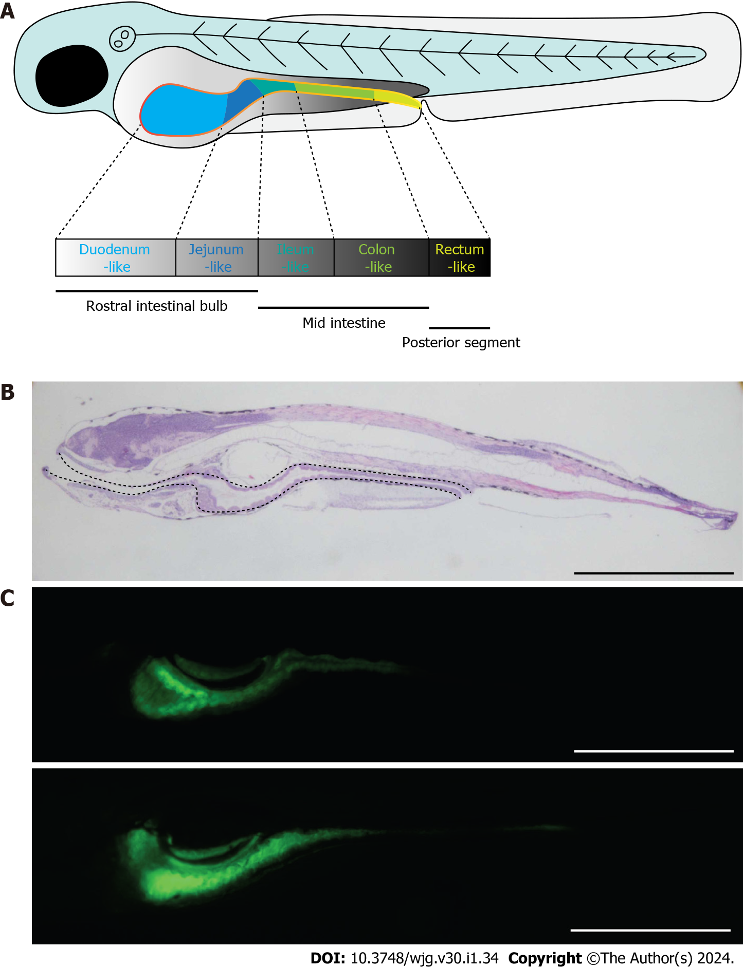
Figure 4 Zebrafish as a good candidate model for the study of Crohn’s disease.
A: Schematic diagram showing the morphology of zebrafish embryo at 5 dpf. The solid yellow line indicates the intestine of the embryo; B: Morphology of zebrafish embryo visualized by hematoxylin and eosin staining of embryonic sections at 5 dpf. Sections were cut along the sagittal plane. The dotted line indicates the whole intestine; C: Fluorescence signals of ET33J1: enhanced green fluorescent protein zebrafish reporter (top) and dichloro-dihydro-fluorescein diacetate treatment (bottom) showing the morphology of the whole intestines at 5 dpf (top) and 7 dpf (bottom). Scale bar: 200 µm. dpf: Days post fertilization.
- Citation: Zhou YW, Ren Y, Lu MM, Xu LL, Cheng WX, Zhang MM, Ding LP, Chen D, Gao JG, Du J, Jin CL, Chen CX, Li YF, Cheng T, Jiang PL, Yang YD, Qian PX, Xu PF, Jin X. Crohn’s disease as the intestinal manifestation of pan-lymphatic dysfunction: An exploratory proposal based on basic and clinical data. World J Gastroenterol 2024; 30(1): 34-49
- URL: https://www.wjgnet.com/1007-9327/full/v30/i1/34.htm
- DOI: https://dx.doi.org/10.3748/wjg.v30.i1.34


