Copyright
©The Author(s) 2024.
World J Gastroenterol. Jan 7, 2024; 30(1): 108-111
Published online Jan 7, 2024. doi: 10.3748/wjg.v30.i1.108
Published online Jan 7, 2024. doi: 10.3748/wjg.v30.i1.108
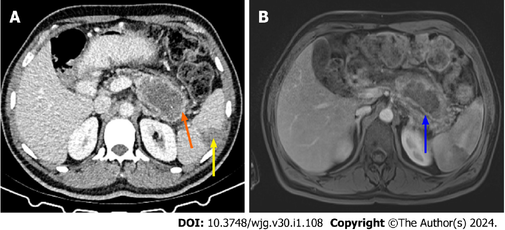
Figure 3 Computed tomography and contrast enhanced magnetic resonance imaging in necrotizing pancreatitis (original image).
A: Computed tomography (CT) image of a patient with known necrotizing pancreatitis. On the pancreatic bed, 73 mm × 45 mm fluid collection with irregular thick walls (orange arrow) was seen along with a focal wedge shaped peripheral non-enhancement of spleen consistent with infarction (yellow arrow); B: Two days following the CT scan, the same patient’s contrast-enhanced T1 weighted fat-saturated magnetic resonance image showed thromboembolic hypointensity in the splenic artery (blue arrow).
- Citation: Ozturk MO, Aydin S. Complementary comments on diagnosis, severity and prognosis prediction of acute pancreatitis. World J Gastroenterol 2024; 30(1): 108-111
- URL: https://www.wjgnet.com/1007-9327/full/v30/i1/108.htm
- DOI: https://dx.doi.org/10.3748/wjg.v30.i1.108


