Copyright
©The Author(s) 2023.
World J Gastroenterol. Mar 7, 2023; 29(9): 1475-1491
Published online Mar 7, 2023. doi: 10.3748/wjg.v29.i9.1475
Published online Mar 7, 2023. doi: 10.3748/wjg.v29.i9.1475
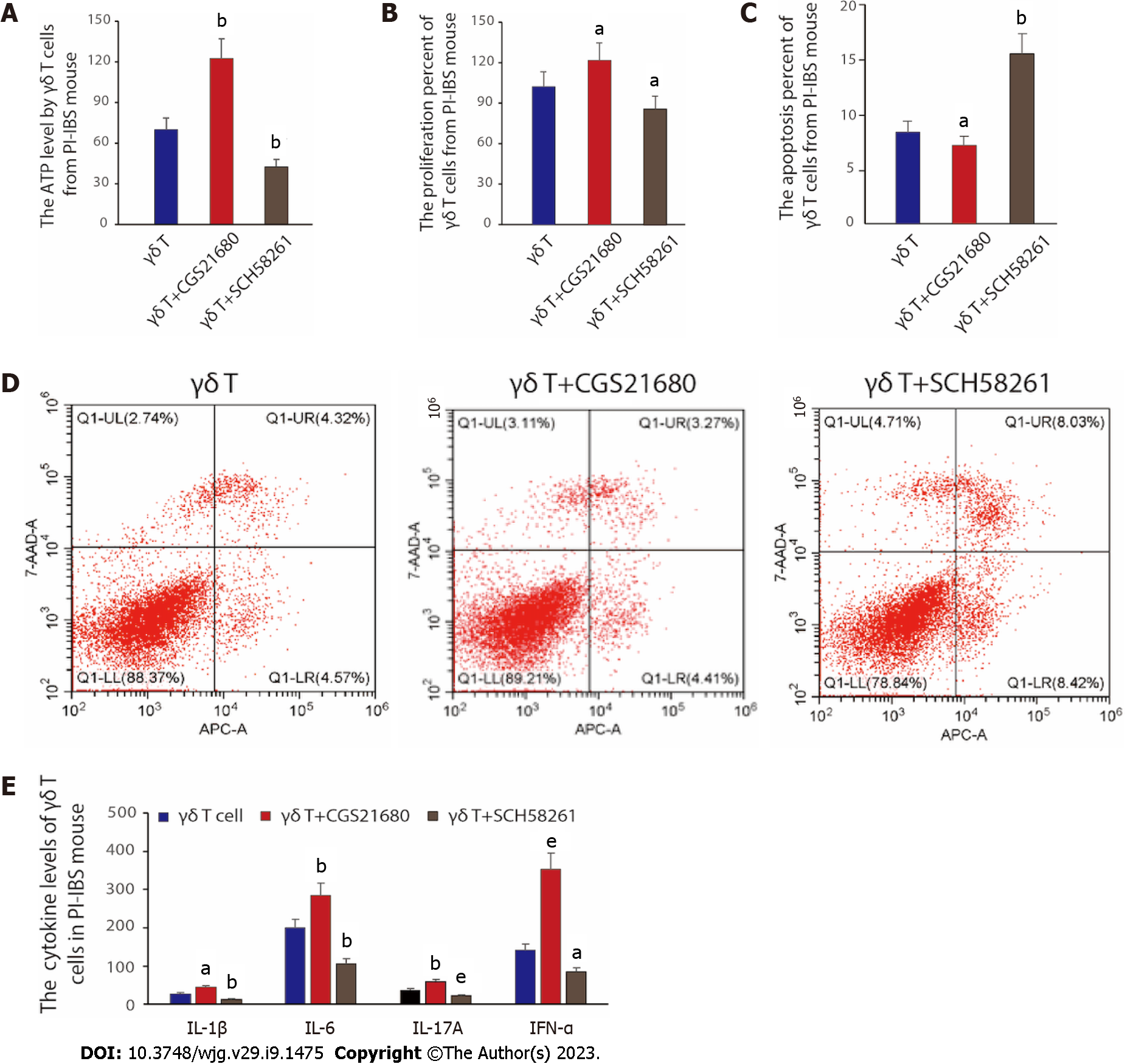
Figure 4 Functional evaluation of γδ T cells in vitro.
A: The ATP level changed by differently treated γδ T cells from post-infectious irritable bowel syndrome (PI-IBS) mice. The data was obtained from six mice in each group; B: The proliferation percent of γδ T cells from PI-IBS mouse was analysed through CCK8 assay. The samples were taken from six mice in each group; C: The apoptosis percent of γδ T cells from PI-IBS mouse measured by fluorescence-activated cell sorting. The γδ T cells were taken from six mice in each group; D: Apoptosis rates were detected according to Annexin V and PI double-staining method; E: The cytokines level of IL-1β, IL-6, IL-17A and IFN-α from PI-IBS mice after reinfusing control γδ T cells and γδ T cells treated with CGS21680 or SCH58261, respectively. The data was obtained from six mice in each group. PI-IBS: Post-infectious irritable bowel syndrome. aP < 0.05, bP < 0.01, eP < 0.001 vs the γδ T cell group.
- Citation: Dong LW, Chen YY, Chen CC, Ma ZC, Fu J, Huang BL, Liu FJ, Liang DC, Sun DM, Lan C. Adenosine 2A receptor contributes to the facilitation of post-infectious irritable bowel syndrome by γδ T cells via the PKA/CREB/NF-κB signaling pathway. World J Gastroenterol 2023; 29(9): 1475-1491
- URL: https://www.wjgnet.com/1007-9327/full/v29/i9/1475.htm
- DOI: https://dx.doi.org/10.3748/wjg.v29.i9.1475


