Copyright
©The Author(s) 2023.
World J Gastroenterol. Feb 21, 2023; 29(7): 1235-1242
Published online Feb 21, 2023. doi: 10.3748/wjg.v29.i7.1235
Published online Feb 21, 2023. doi: 10.3748/wjg.v29.i7.1235
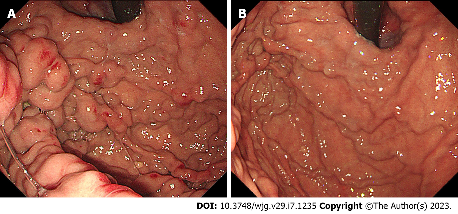
Figure 3 Endoscopic image on admission and 1 mo later.
A: Endoscopic image before partial splenic embolization (PSE) showing diffused gastric varices; B: Endoscopic image after PSE showing alleviation of gastric varices.
- Citation: Wang GC, Huang GJ, Zhang CQ, Ding Q. Percutaneous transhepatic intraportal biopsy using gastroscope biopsy forceps for diagnosis of a pancreatic neuroendocrine neoplasm: A case report. World J Gastroenterol 2023; 29(7): 1235-1242
- URL: https://www.wjgnet.com/1007-9327/full/v29/i7/1235.htm
- DOI: https://dx.doi.org/10.3748/wjg.v29.i7.1235


