Copyright
©The Author(s) 2023.
World J Gastroenterol. Feb 7, 2023; 29(5): 834-850
Published online Feb 7, 2023. doi: 10.3748/wjg.v29.i5.834
Published online Feb 7, 2023. doi: 10.3748/wjg.v29.i5.834
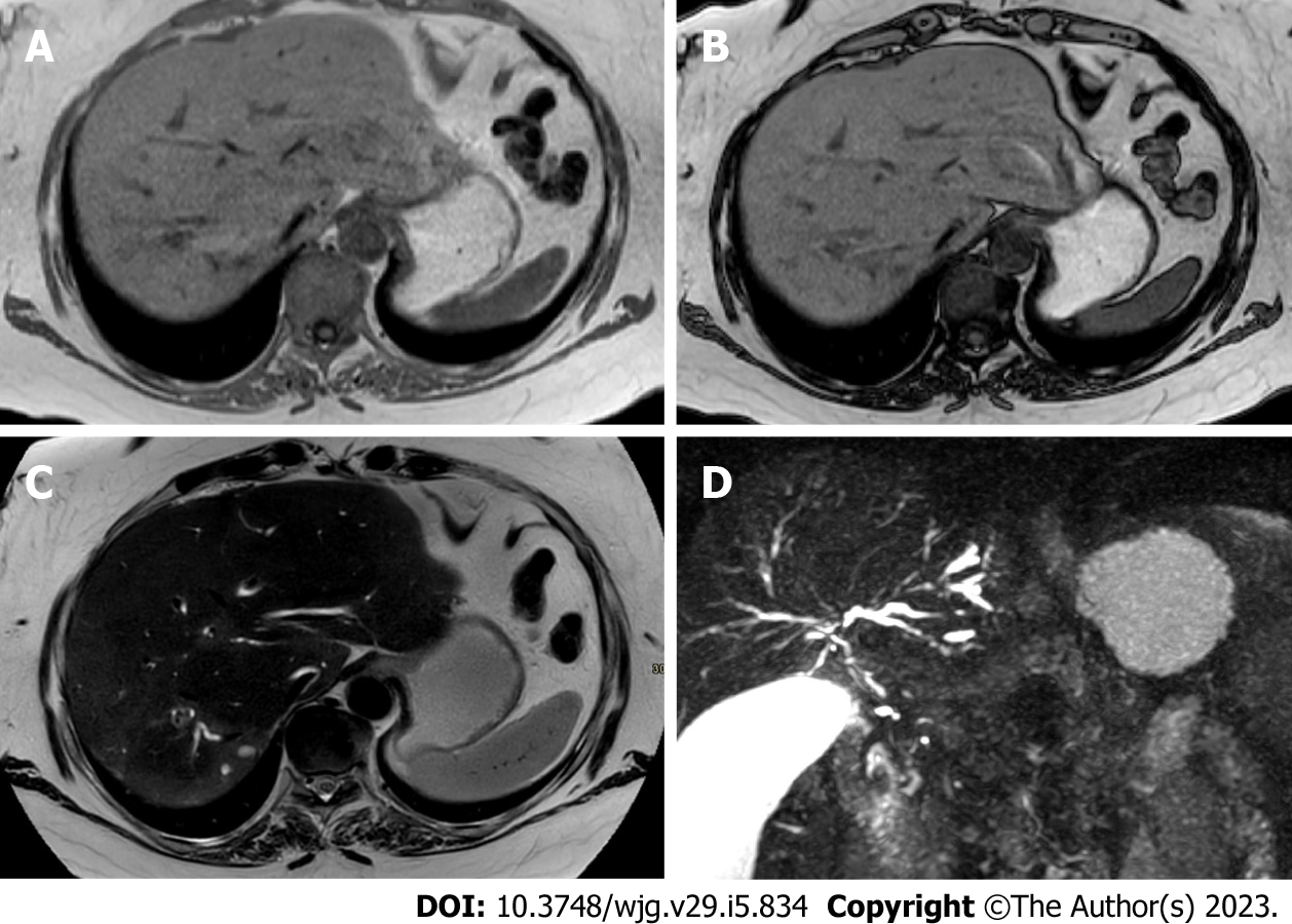
Figure 8 A 44-year-old woman with coronavirus disease 2019 infection underwent magnetic resonance cholangiopancreatography due to elevated biliary enzymes with negative findings on ultrasonography and contrast-enhanced computed tomography.
A and B: In-phase (A) and out-of-phase (B) imaging demonstrates hepatic homogeneous parenchyma without areas of fatty infiltration; C: On the T2-weighted image liver inflammation, especially in the right lobe, is characterized by areas of slight hyperenhancement, located nearby the biliary ducts; D: Finally, magnetic resonance cholangiopancreatography allows the evaluation of the biliary tree, showing multiple and diffuse focal biliary stenosis, in particular in the right lobe, with upstream dilation of small biliary ducts. This radiological aspect is typical of sclerosing cholangitis and the final diagnosis was a biliary involvement due to severe acute respiratory syndrome coronavirus 2.
- Citation: Ippolito D, Maino C, Vernuccio F, Cannella R, Inchingolo R, Dezio M, Faletti R, Bonaffini PA, Gatti M, Sironi S. Liver involvement in patients with COVID-19 infection: A comprehensive overview of diagnostic imaging features. World J Gastroenterol 2023; 29(5): 834-850
- URL: https://www.wjgnet.com/1007-9327/full/v29/i5/834.htm
- DOI: https://dx.doi.org/10.3748/wjg.v29.i5.834


