Copyright
©The Author(s) 2023.
World J Gastroenterol. Feb 7, 2023; 29(5): 834-850
Published online Feb 7, 2023. doi: 10.3748/wjg.v29.i5.834
Published online Feb 7, 2023. doi: 10.3748/wjg.v29.i5.834
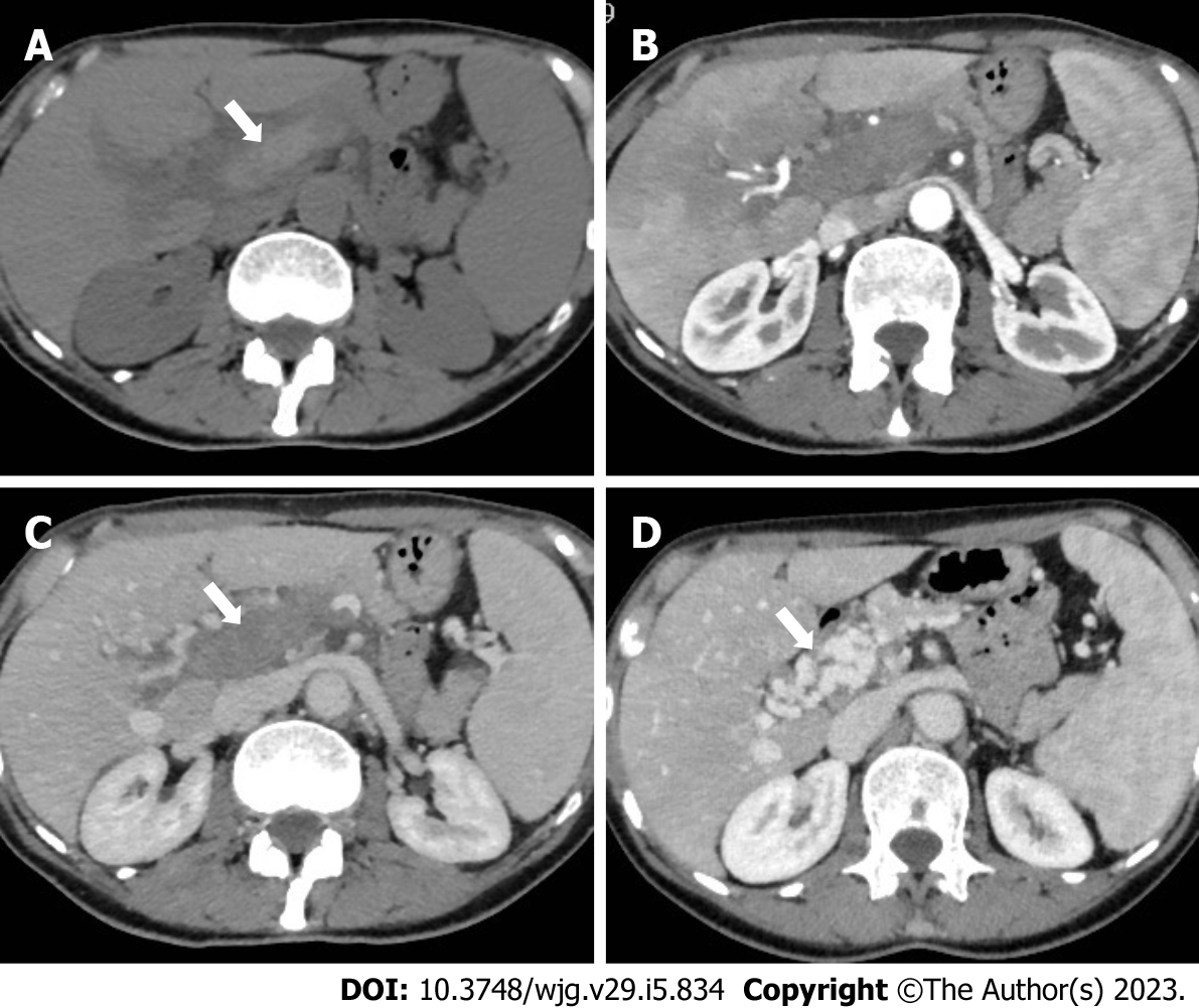
Figure 5 A 62-year-old woman with acute portal vein thrombosis after coronavirus disease 2019 infection.
A-C: Computed tomography (CT) images show acute thrombosis with hyperintense thrombus on unenhanced phase (A, arrow), heterogeneous enhancement of the liver parenchyma on hepatic arterial phase (B), and complete portal vein thrombosis on portal venous phase (C, arrow); D: Contrast-enhanced CT at 6-mo follow-up demonstrates chronic findings of portal cavernoma with multiple collateral vessels at the hepatic hilum (arrow).
- Citation: Ippolito D, Maino C, Vernuccio F, Cannella R, Inchingolo R, Dezio M, Faletti R, Bonaffini PA, Gatti M, Sironi S. Liver involvement in patients with COVID-19 infection: A comprehensive overview of diagnostic imaging features. World J Gastroenterol 2023; 29(5): 834-850
- URL: https://www.wjgnet.com/1007-9327/full/v29/i5/834.htm
- DOI: https://dx.doi.org/10.3748/wjg.v29.i5.834


