Copyright
©The Author(s) 2023.
World J Gastroenterol. Feb 7, 2023; 29(5): 834-850
Published online Feb 7, 2023. doi: 10.3748/wjg.v29.i5.834
Published online Feb 7, 2023. doi: 10.3748/wjg.v29.i5.834
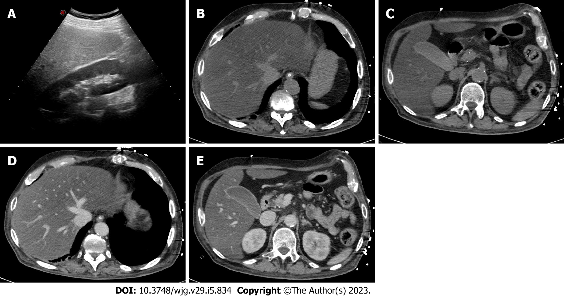
Figure 2 A 45-year-old woman with coronavirus disease 2019 infection underwent abdominal ultrasonography due to elevated liver enzymes.
A: As shown on ultrasonography, the liver is hyperechoic in comparison to the renal parenchyma, suggesting marked steatosis; B-E: The patient underwent contrast-enhanced computed tomography; B and C: On unenhanced images, the liver is diffusely hypoattenuating while its main vessels (i.e. portal vein and hepatic veins) appear brighter than the parenchyma due to a marked hepatic fatty infiltration; D and E: On post-contrast images, the liver is homogeneous without any focal lesion; C and E: The gallbladder is filled by sludge, with normal wall thickness. Finally, the combination of clinical and radiological findings allowed the final diagnosis of steatohepatitis.
- Citation: Ippolito D, Maino C, Vernuccio F, Cannella R, Inchingolo R, Dezio M, Faletti R, Bonaffini PA, Gatti M, Sironi S. Liver involvement in patients with COVID-19 infection: A comprehensive overview of diagnostic imaging features. World J Gastroenterol 2023; 29(5): 834-850
- URL: https://www.wjgnet.com/1007-9327/full/v29/i5/834.htm
- DOI: https://dx.doi.org/10.3748/wjg.v29.i5.834


