Copyright
©The Author(s) 2023.
World J Gastroenterol. Dec 14, 2023; 29(46): 6049-6059
Published online Dec 14, 2023. doi: 10.3748/wjg.v29.i46.6049
Published online Dec 14, 2023. doi: 10.3748/wjg.v29.i46.6049
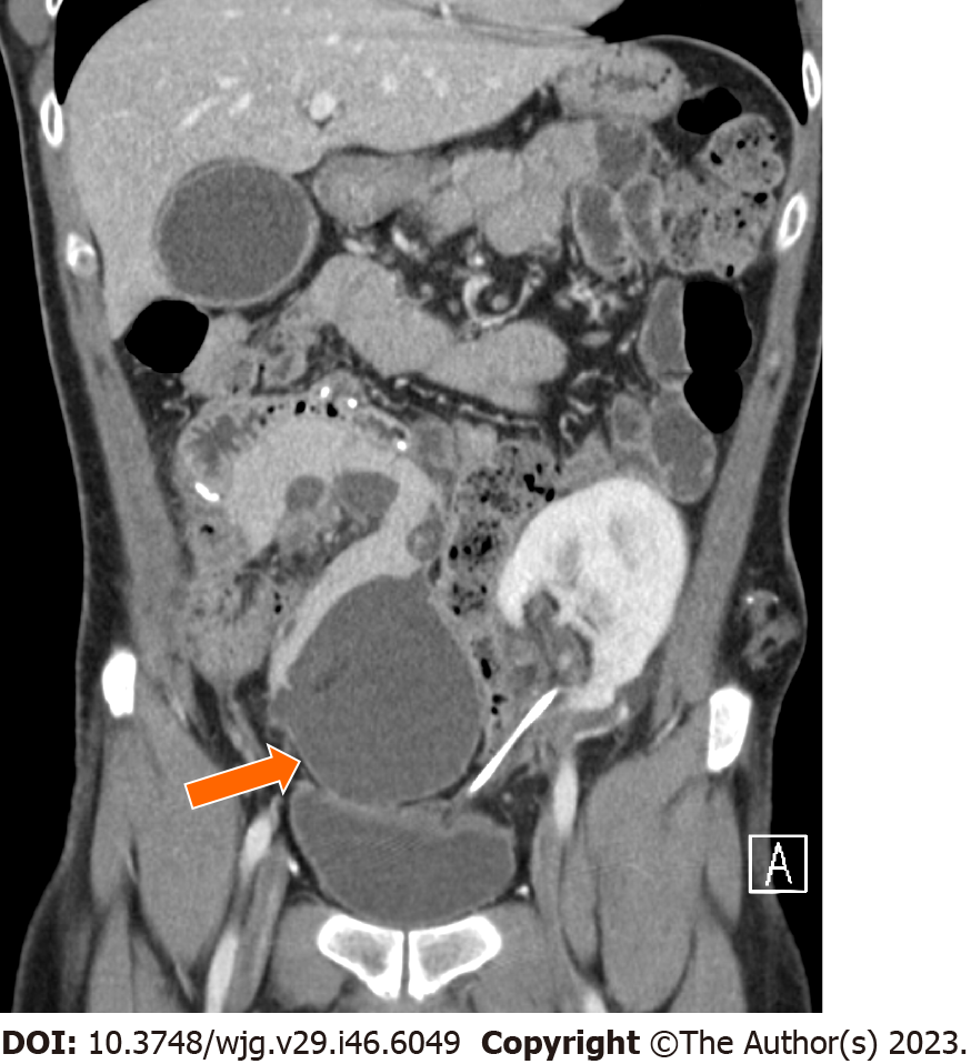
Figure 8 A 35-year-old man after four weeks from edematous pancreatitis.
Contrast enhanced computed tomography in the coronal plane shows an encapsulated fluid collection (arrow) of homogeneously low attenuation surrounded by a well-defined enhancing wall consistent with pancreatic pseudocyst.
- Citation: D'Alessandro C, Todisco M, Di Bella C, Crimì F, Furian L, Quaia E, Vernuccio F. Surgical complications after pancreatic transplantation: A computed tomography imaging pictorial review. World J Gastroenterol 2023; 29(46): 6049-6059
- URL: https://www.wjgnet.com/1007-9327/full/v29/i46/6049.htm
- DOI: https://dx.doi.org/10.3748/wjg.v29.i46.6049


