Copyright
©The Author(s) 2023.
World J Gastroenterol. Nov 28, 2023; 29(44): 5935-5944
Published online Nov 28, 2023. doi: 10.3748/wjg.v29.i44.5935
Published online Nov 28, 2023. doi: 10.3748/wjg.v29.i44.5935
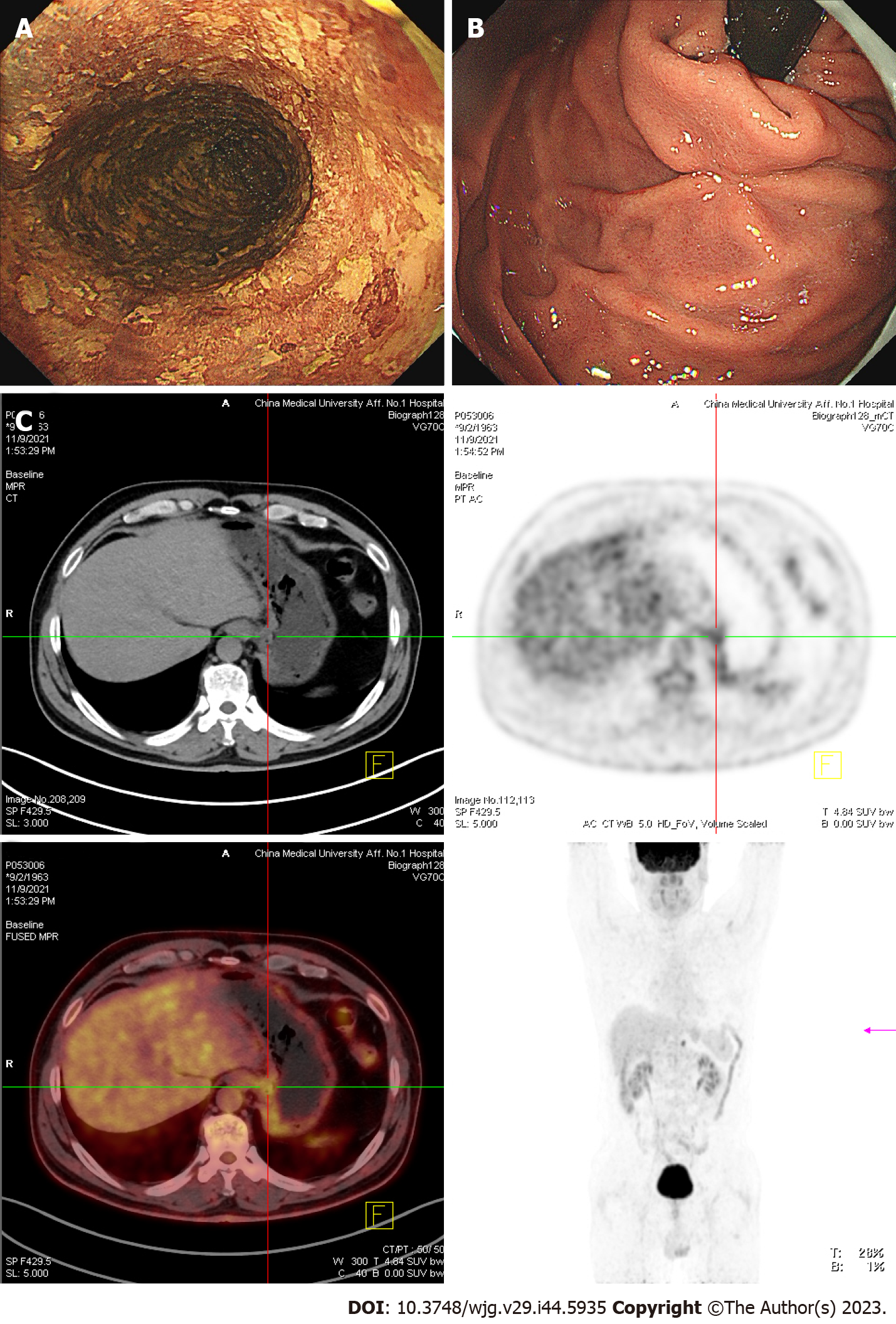
Figure 4 In November 2021, the patient underwent the third re-examination 15 mo after the first endoscopic submucosal dissection.
A: The entire esophagus showed patchy, lightly stained areas after iodine staining in gastroendoscopy. B: The mucosa of the gastric fundus and cardia was smooth except for the scar after Endoscopic submucosal dissection operation, and no abnormality was noted on gastroendoscopy. C: Computed tomography (CT) revealed no abnormal thickening in the gastrointestinal wall. However, positron emission tomography-CT revealed that the metabolism of the lower esophagus and stomach cardia wall slightly increased. A lymph node was noted in the retroperitoneal area with high-density shadow and increased metabolism.
- Citation: Yang MQ, Sun MJ, Zhang HJ. Mucosal esophageal carcinoma following endoscopic submucosal dissection with giant gastric metastasis: A case report and review of literature. World J Gastroenterol 2023; 29(44): 5935-5944
- URL: https://www.wjgnet.com/1007-9327/full/v29/i44/5935.htm
- DOI: https://dx.doi.org/10.3748/wjg.v29.i44.5935


