Copyright
©The Author(s) 2023.
World J Gastroenterol. Nov 28, 2023; 29(44): 5935-5944
Published online Nov 28, 2023. doi: 10.3748/wjg.v29.i44.5935
Published online Nov 28, 2023. doi: 10.3748/wjg.v29.i44.5935
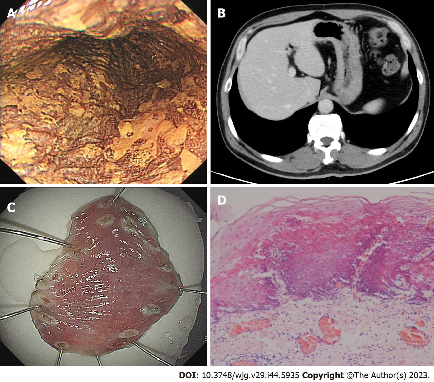
Figure 1 The first gastroendoscopy examination in August 2020.
A: After iodine staining, the esophageal mucosa was scattered in patchy light-stained areas, ranging from 0.2 cm to 0.4 cm, and a 2.0 cm × 1.0 cm, superficial, flat lesion (30 cm from the incisor teeth) did not stain, whereas the pink areas were positive; B: Computed tomography showed no abnormal thickening in the gastrointestinal wall; C: Endoscopic submucosal dissection was performed 1 wk after gastroendoscopy; D: Postoperative pathology revealed moderate to severe dysplasia of squamous epithelium.
- Citation: Yang MQ, Sun MJ, Zhang HJ. Mucosal esophageal carcinoma following endoscopic submucosal dissection with giant gastric metastasis: A case report and review of literature. World J Gastroenterol 2023; 29(44): 5935-5944
- URL: https://www.wjgnet.com/1007-9327/full/v29/i44/5935.htm
- DOI: https://dx.doi.org/10.3748/wjg.v29.i44.5935


