Copyright
©The Author(s) 2023.
World J Gastroenterol. Oct 28, 2023; 29(40): 5566-5581
Published online Oct 28, 2023. doi: 10.3748/wjg.v29.i40.5566
Published online Oct 28, 2023. doi: 10.3748/wjg.v29.i40.5566
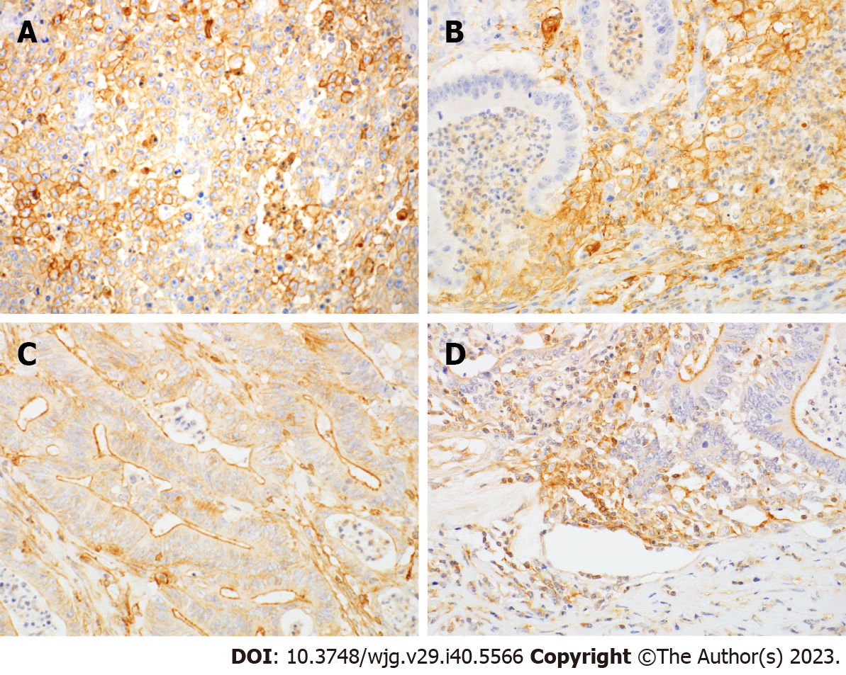
Figure 1 Immunohistochemical localization of programmed cell death-ligand 1 and programmed cell death-ligand 2 in small bowel adenocarcinoma.
Magnification 400×. A: T-programmed cell death-ligand 1 expression was membranous and cytoplasmic; B: I-programmed cell death-ligand 1 (I-PD-L1) was positive in peritumoral lymphocytes and macrophages; C: The predominant pattern of T-programmed cell death-ligand 2 expression was in the apical membrane; D: I-programmed cell death-ligand 2 expression was positive in peritumoral lymphocytes and macrophages, similar to that of I-PD-L1.
- Citation: Hoshimoto A, Tatsuguchi A, Hamakubo R, Nishimoto T, Omori J, Akimoto N, Tanaka S, Fujimori S, Hatori T, Shimizu A, Iwakiri K. Clinical significance of programmed cell death-ligand expression in small bowel adenocarcinoma is determined by the tumor microenvironment. World J Gastroenterol 2023; 29(40): 5566-5581
- URL: https://www.wjgnet.com/1007-9327/full/v29/i40/5566.htm
- DOI: https://dx.doi.org/10.3748/wjg.v29.i40.5566


