Copyright
©The Author(s) 2023.
World J Gastroenterol. Sep 28, 2023; 29(36): 5180-5197
Published online Sep 28, 2023. doi: 10.3748/wjg.v29.i36.5180
Published online Sep 28, 2023. doi: 10.3748/wjg.v29.i36.5180
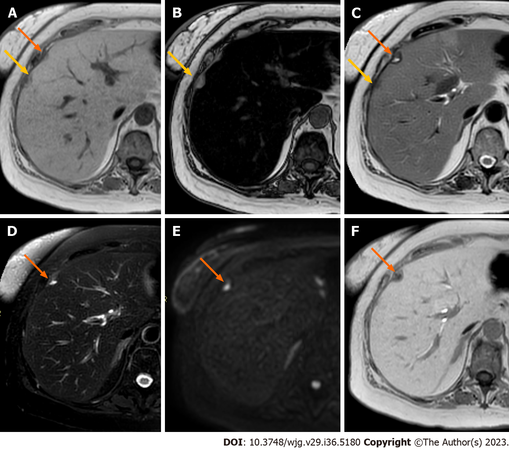
Figure 7 Liver metastases from pancreatic adenocarcinoma.
A: EOB-magnetic resonance imaging (MRI) of a 64-year-old woman shows two adjacent focal liver lesions (yellow and orange arrows) in the subcapsular region on the in-phase imaging; B: On out-phase imaging, one lesion (yellow arrow) shows hyperintensity, while the other one shows slight hypointensity. The liver is characterized by severe steatosis, considering the important signal drop-off between in-phase and out-of-phase imaging; C: On T2-weighted (T2W) imaging, one lesion (yellow arrow) is hypointense, while the other one (orange arrow) demonstrates inhomogeneous hyperintense signal; D: On fat-sat T2W sequence the low signal of the first lesion and the inhomogeneous high signal of the second one (orange arrow) are confirmed; E: Diffusion weighted imaging reveals focal restriction of diffusion corresponding to only one of the two lesions (orange arrow); F: On the hepatobiliary phase one lesion is isointense to liver parenchyma, while the second lesion shows a hypointense aspect. The final diagnosis for the lesion tagged by the orange arrow was liver metastasis from pancreatic adenocarcinoma, while the MRI characteristics of the lesion tagged by the yellow arrow are consistent with a focal fatty-sparing area.
- Citation: Maino C, Vernuccio F, Cannella R, Cortese F, Franco PN, Gaetani C, Giannini V, Inchingolo R, Ippolito D, Defeudis A, Pilato G, Tore D, Faletti R, Gatti M. Liver metastases: The role of magnetic resonance imaging. World J Gastroenterol 2023; 29(36): 5180-5197
- URL: https://www.wjgnet.com/1007-9327/full/v29/i36/5180.htm
- DOI: https://dx.doi.org/10.3748/wjg.v29.i36.5180


