Copyright
©The Author(s) 2023.
World J Gastroenterol. Sep 28, 2023; 29(36): 5180-5197
Published online Sep 28, 2023. doi: 10.3748/wjg.v29.i36.5180
Published online Sep 28, 2023. doi: 10.3748/wjg.v29.i36.5180
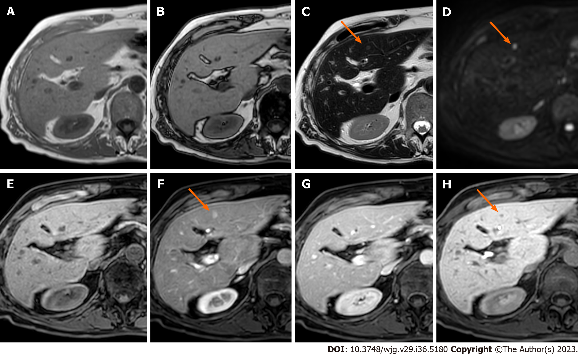
Figure 6 Liver metastases from ileal neuroendocrine tumor.
A: EOB-magnetic resonance imaging of a 55-year-old female patient shows a focal liver lesion isointense on in-phase images; B: On out-of-phase images, the lesions persist isointense compared to the healthy liver parenchyma; C: On T2-weighted images the lesion appears slightly hyperintense (orange arrow); D: Diffusion weighted imaging reveal restriction of diffusion of the lesion (orange arrow); E: Before contrast administration the lesion is isointense; F: During the post-contrast late hepatic arterial phase the lesion appears hypervascular (orange arrow); G: The lesion is isointense on the portal-venous phase; H: On the hepatobiliary phase lesion’s low signal intensity is observed (orange arrow).
- Citation: Maino C, Vernuccio F, Cannella R, Cortese F, Franco PN, Gaetani C, Giannini V, Inchingolo R, Ippolito D, Defeudis A, Pilato G, Tore D, Faletti R, Gatti M. Liver metastases: The role of magnetic resonance imaging. World J Gastroenterol 2023; 29(36): 5180-5197
- URL: https://www.wjgnet.com/1007-9327/full/v29/i36/5180.htm
- DOI: https://dx.doi.org/10.3748/wjg.v29.i36.5180


