Copyright
©The Author(s) 2023.
World J Gastroenterol. Sep 28, 2023; 29(36): 5180-5197
Published online Sep 28, 2023. doi: 10.3748/wjg.v29.i36.5180
Published online Sep 28, 2023. doi: 10.3748/wjg.v29.i36.5180
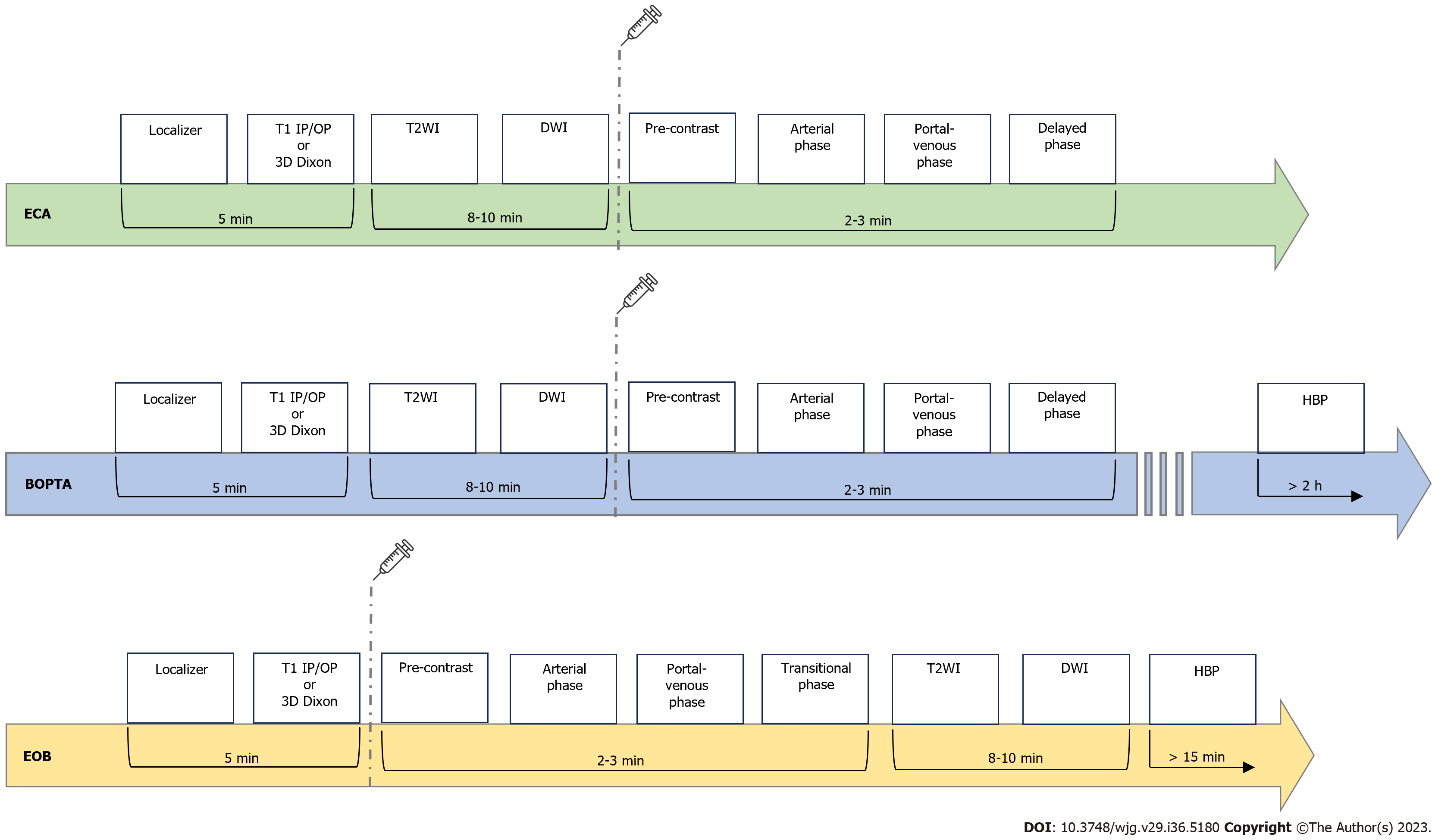
Figure 1 Schematic summary of magnetic resonance imaging protocols by using ECAs and hepatobiliary contrast agents (Gd-BOPTA and Gd-EOB-DTPA).
ECA: Extra-cellular; BOPTA: Gadobenate dimeglumine; EOB: Gadoxetic acid, disodium; T1 IP: T1-weighted in-phase imaging; T1 OP: T1-weighted out-of-phase imaging; DWI: Diffusion weighted imaging; HBP: Hepatobiliary phase; T2WI: T2-weighted imaging; HBP: Hepatobiliary phase.
- Citation: Maino C, Vernuccio F, Cannella R, Cortese F, Franco PN, Gaetani C, Giannini V, Inchingolo R, Ippolito D, Defeudis A, Pilato G, Tore D, Faletti R, Gatti M. Liver metastases: The role of magnetic resonance imaging. World J Gastroenterol 2023; 29(36): 5180-5197
- URL: https://www.wjgnet.com/1007-9327/full/v29/i36/5180.htm
- DOI: https://dx.doi.org/10.3748/wjg.v29.i36.5180


