Copyright
©The Author(s) 2023.
World J Gastroenterol. Sep 7, 2023; 29(33): 5005-5013
Published online Sep 7, 2023. doi: 10.3748/wjg.v29.i33.5005
Published online Sep 7, 2023. doi: 10.3748/wjg.v29.i33.5005
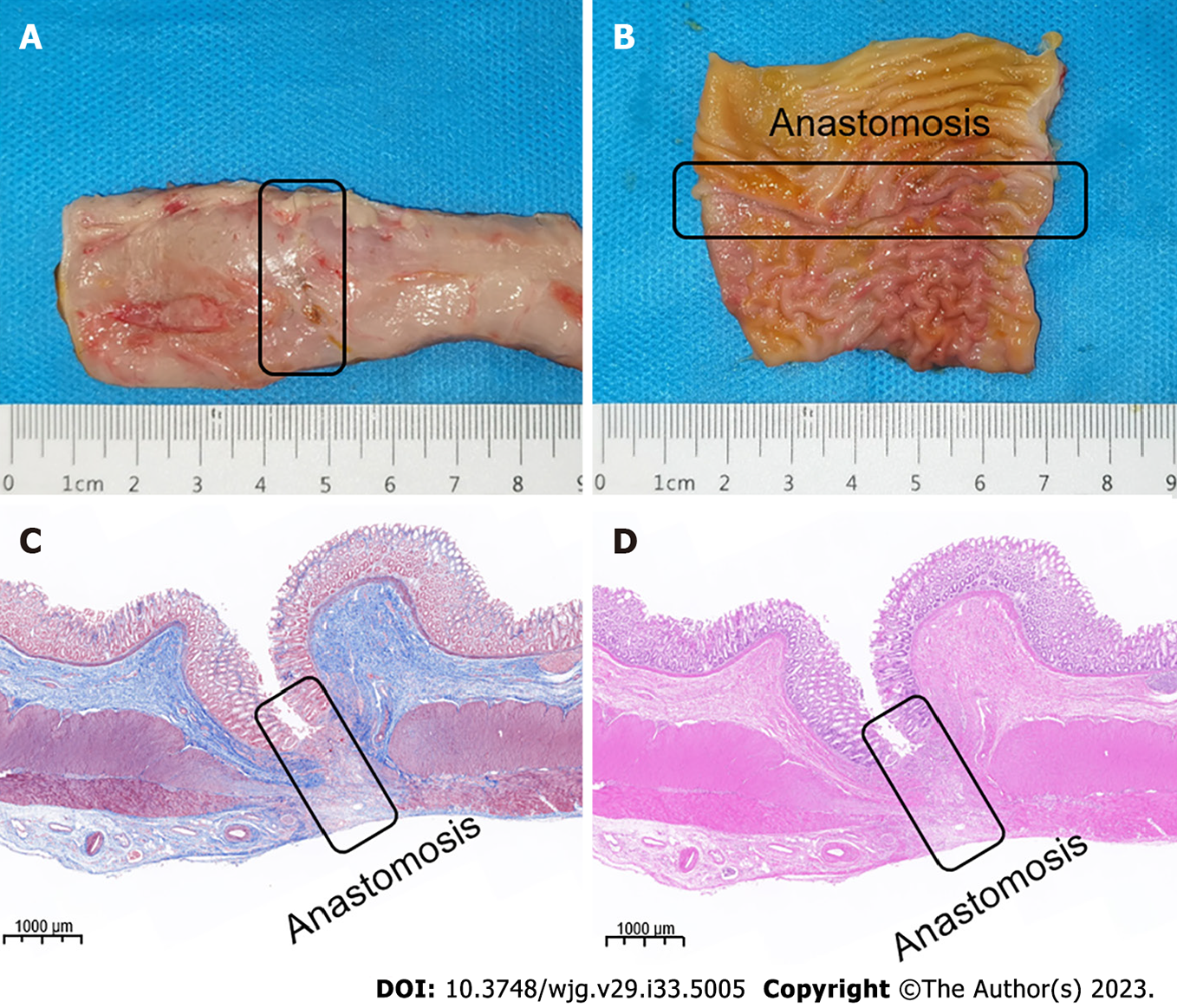
Figure 5 Magnetic compression anastomosis specimen.
A: Serous surface of the anastomosis; B: Colonic anastomosis seen on mucosal surface; C: Masson’s staining of anastomosis; D: Hematoxylin and eosin staining of anastomosis.
- Citation: Zhang MM, Zhao GB, Zhang HZ, Xu SQ, Shi AH, Mao JQ, Gai JC, Zhang YH, Ma J, Li Y, Lyu Y, Yan XP. Novel deformable self-assembled magnetic anastomosis ring for endoscopic treatment of colonic stenosis via natural orifice. World J Gastroenterol 2023; 29(33): 5005-5013
- URL: https://www.wjgnet.com/1007-9327/full/v29/i33/5005.htm
- DOI: https://dx.doi.org/10.3748/wjg.v29.i33.5005


