Copyright
©The Author(s) 2023.
World J Gastroenterol. Jan 21, 2023; 29(3): 521-535
Published online Jan 21, 2023. doi: 10.3748/wjg.v29.i3.521
Published online Jan 21, 2023. doi: 10.3748/wjg.v29.i3.521
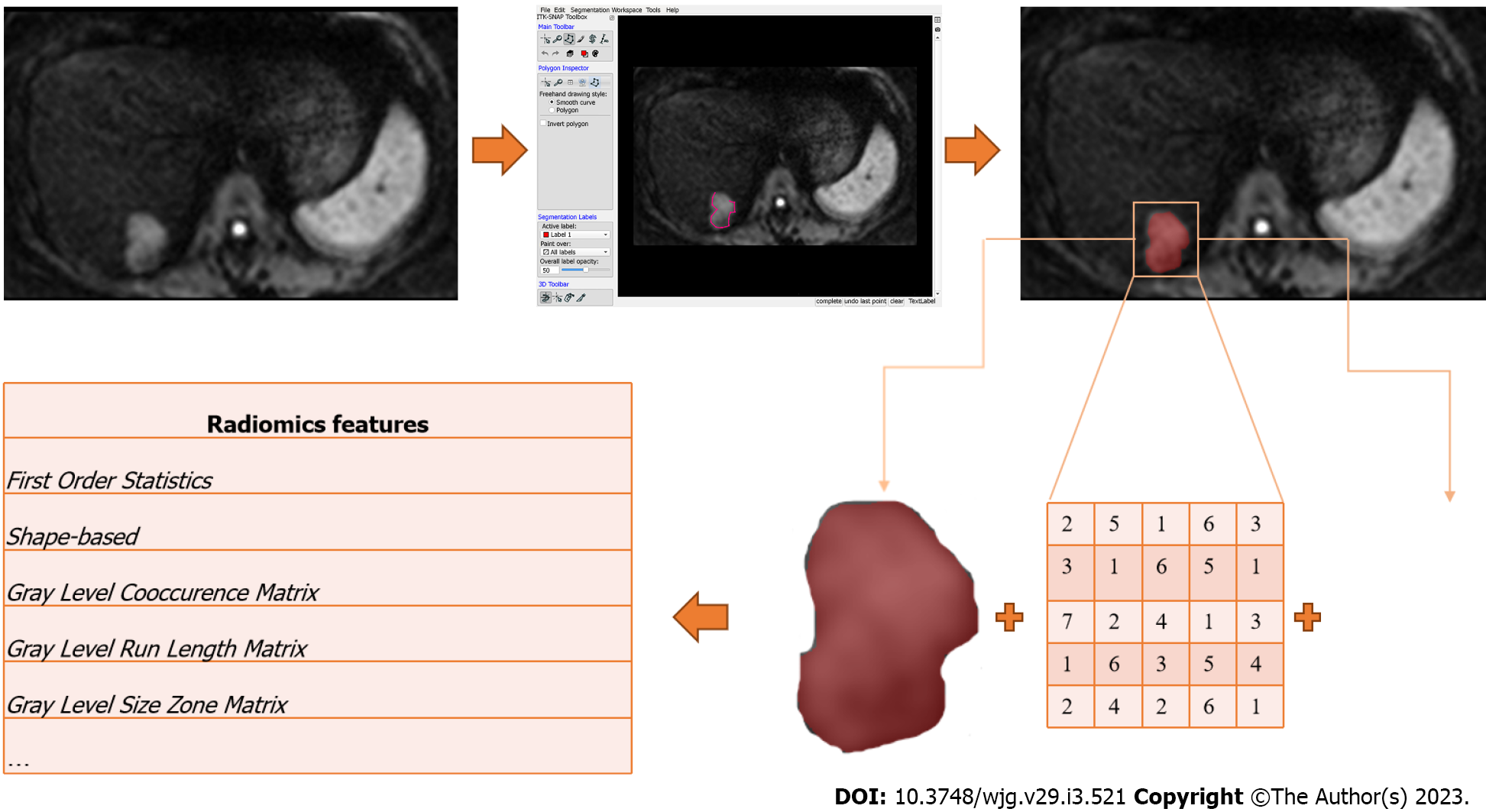
Figure 2 Schematic diagram showing how radiomics features can be extracted from medical images using a diffusion-weighted imaging image from an magnetic resonance imaging scan of a patient with colorectal liver metastasis as an example.
The process begins on the left upper corner with image acquisition, followed by lesion segmentation on a dedicated software leading to a region of interest. The shape of the region of interest as well as the distribution and spatial relation of intensity values of each pixel are computationally analysed to extract radiomics features of different order.
- Citation: Caruso M, Stanzione A, Prinster A, Pizzuti LM, Brunetti A, Maurea S, Mainenti PP. Role of advanced imaging techniques in the evaluation of oncological therapies in patients with colorectal liver metastases. World J Gastroenterol 2023; 29(3): 521-535
- URL: https://www.wjgnet.com/1007-9327/full/v29/i3/521.htm
- DOI: https://dx.doi.org/10.3748/wjg.v29.i3.521


