Copyright
©The Author(s) 2023.
World J Gastroenterol. Jul 21, 2023; 29(27): 4222-4235
Published online Jul 21, 2023. doi: 10.3748/wjg.v29.i27.4222
Published online Jul 21, 2023. doi: 10.3748/wjg.v29.i27.4222
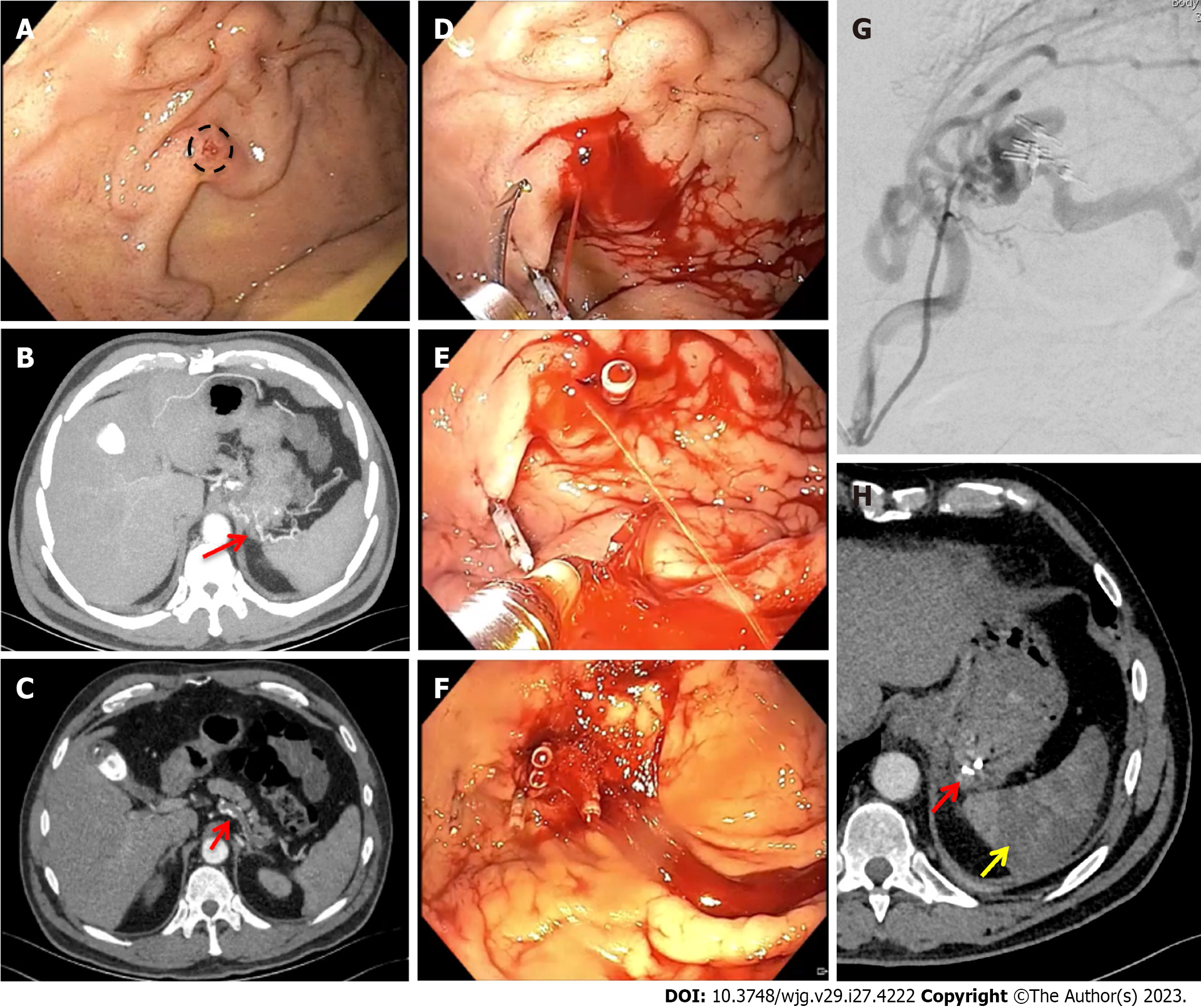
Figure 3 Non-variceal upper gastrointestinal bleeding due to gastric submucosal arterial collaterals secondary to splenic artery thrombosis.
A: Retroflexed endoscopic view of the gastric fundus showing varicose-shaped submucosal vessels with a small erosion (circle), in the absence of active bleeding; B and C: Pre-operative axial computed tomography arterial phase showing an arterial cluster at the gastric fundus (B: arrow) arising from splenic artery collateral vessels due to splenic artery complete thrombosis (C: arrow); D-F: Successful endoscopic mechanical hemostasis attempt, with intraprocedural spurting active bleeding following first endoclip application (D and E); G: Operative angiography with selective arterial embolization using microspheres; H: Post-operative axial computed tomography arterial phase showing gastric vascular lesion exclusion (red arrow) and splenic infarction (yellow arrow).
- Citation: Martino A, Di Serafino M, Orsini L, Giurazza F, Fiorentino R, Crolla E, Campione S, Molino C, Romano L, Lombardi G. Rare causes of acute non-variceal upper gastrointestinal bleeding: A comprehensive review. World J Gastroenterol 2023; 29(27): 4222-4235
- URL: https://www.wjgnet.com/1007-9327/full/v29/i27/4222.htm
- DOI: https://dx.doi.org/10.3748/wjg.v29.i27.4222


