Copyright
©The Author(s) 2023.
World J Gastroenterol. Jul 14, 2023; 29(26): 4214-4221
Published online Jul 14, 2023. doi: 10.3748/wjg.v29.i26.4214
Published online Jul 14, 2023. doi: 10.3748/wjg.v29.i26.4214
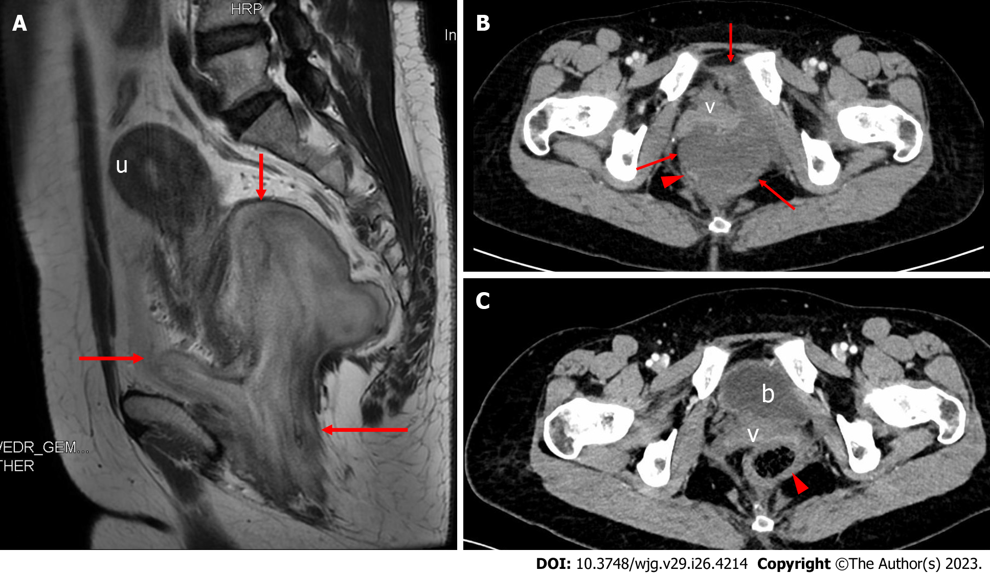
Figure 1 Magnetic resonance imaging and contrast-enhanced computed tomography images in our case.
A: Preoperative sagittal T2-weighted imaging. The mass (arrows) was 9.5 cm × 8.1 cm in size, showing a layered appearance with alternating high and low signals and a clearly demarcated boundary with respect to surrounding tissue, and the uterus was pushed and displaced cephalad; B: Preoperative transverse contrast-enhanced computed tomography (CECT) showed that the anterior vagina and the right posterior rectum (arrowhead) were pushed by the mass (arrows); C: Six months after surgery, transverse CECT showed that the filled bladder, vagina, and rectum (arrowhead) were in normal positions, with no evidence of tumor recurrence. u: Uterus; v: Vagina; b: Bladder.
- Citation: Zhang Q, Yan HL, Lu Q, Luo Y. Value of contrast-enhanced ultrasound in deep angiomyxoma using a biplane transrectal probe: A case report. World J Gastroenterol 2023; 29(26): 4214-4221
- URL: https://www.wjgnet.com/1007-9327/full/v29/i26/4214.htm
- DOI: https://dx.doi.org/10.3748/wjg.v29.i26.4214


