Copyright
©The Author(s) 2023.
World J Gastroenterol. Jun 21, 2023; 29(23): 3606-3621
Published online Jun 21, 2023. doi: 10.3748/wjg.v29.i23.3606
Published online Jun 21, 2023. doi: 10.3748/wjg.v29.i23.3606
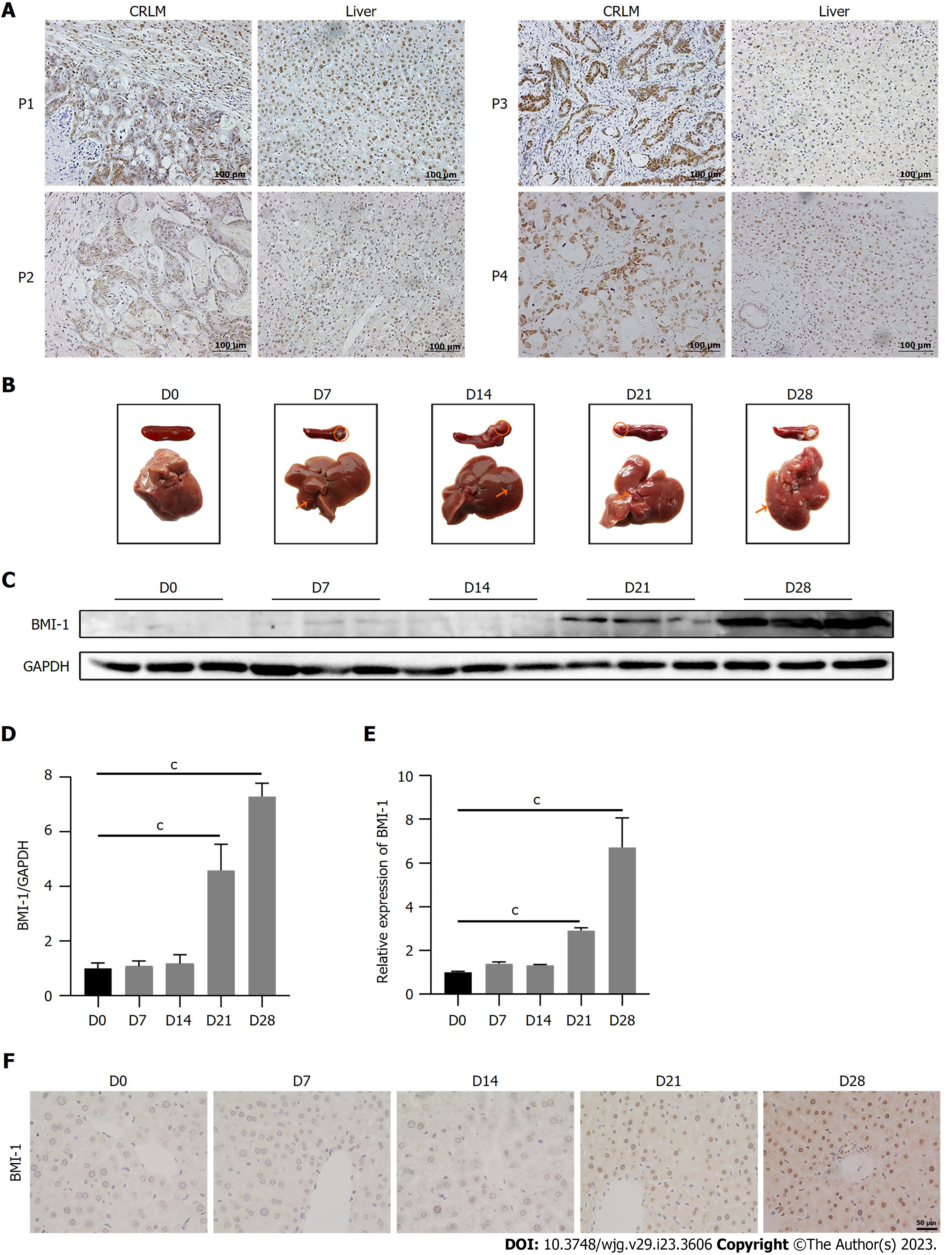
Figure 1 BMI-1 expression increases in liver cells during colorectal cancer liver metastasis.
A: Immunohistochemistry analysis of BMI-1 in liver metastasis and paired liver tissues of colorectal cancer liver metastasis (CRLM) patients; B: Spleens and livers of mice were photographed after intrasplenic injection of CT26 cells at the indicated times; C: Western blot analysis of BMI-1 expression in mouse livers (n = 3); D: Quantitative protein expression of BMI-1 normalized to glyceraldehyde-3-phosphate dehydrogenase (GAPDH) in mouse livers (n = 3); E: Quantitative polymerase chain reaction detection of BMI-1 expression in mouse livers (n = 3); cP < 0.001 vs D0; F: Immunohistochemistry confirmed that BMI-1 expression was increased in mouse livers during CRLM.
- Citation: Jiang ZY, Ma XM, Luan XH, Liuyang ZY, Hong YY, Dai Y, Dong QH, Wang GY. BMI-1 activates hepatic stellate cells to promote the epithelial-mesenchymal transition of colorectal cancer cells. World J Gastroenterol 2023; 29(23): 3606-3621
- URL: https://www.wjgnet.com/1007-9327/full/v29/i23/3606.htm
- DOI: https://dx.doi.org/10.3748/wjg.v29.i23.3606


