Copyright
©The Author(s) 2023.
World J Gastroenterol. Jun 14, 2023; 29(22): 3440-3468
Published online Jun 14, 2023. doi: 10.3748/wjg.v29.i22.3440
Published online Jun 14, 2023. doi: 10.3748/wjg.v29.i22.3440
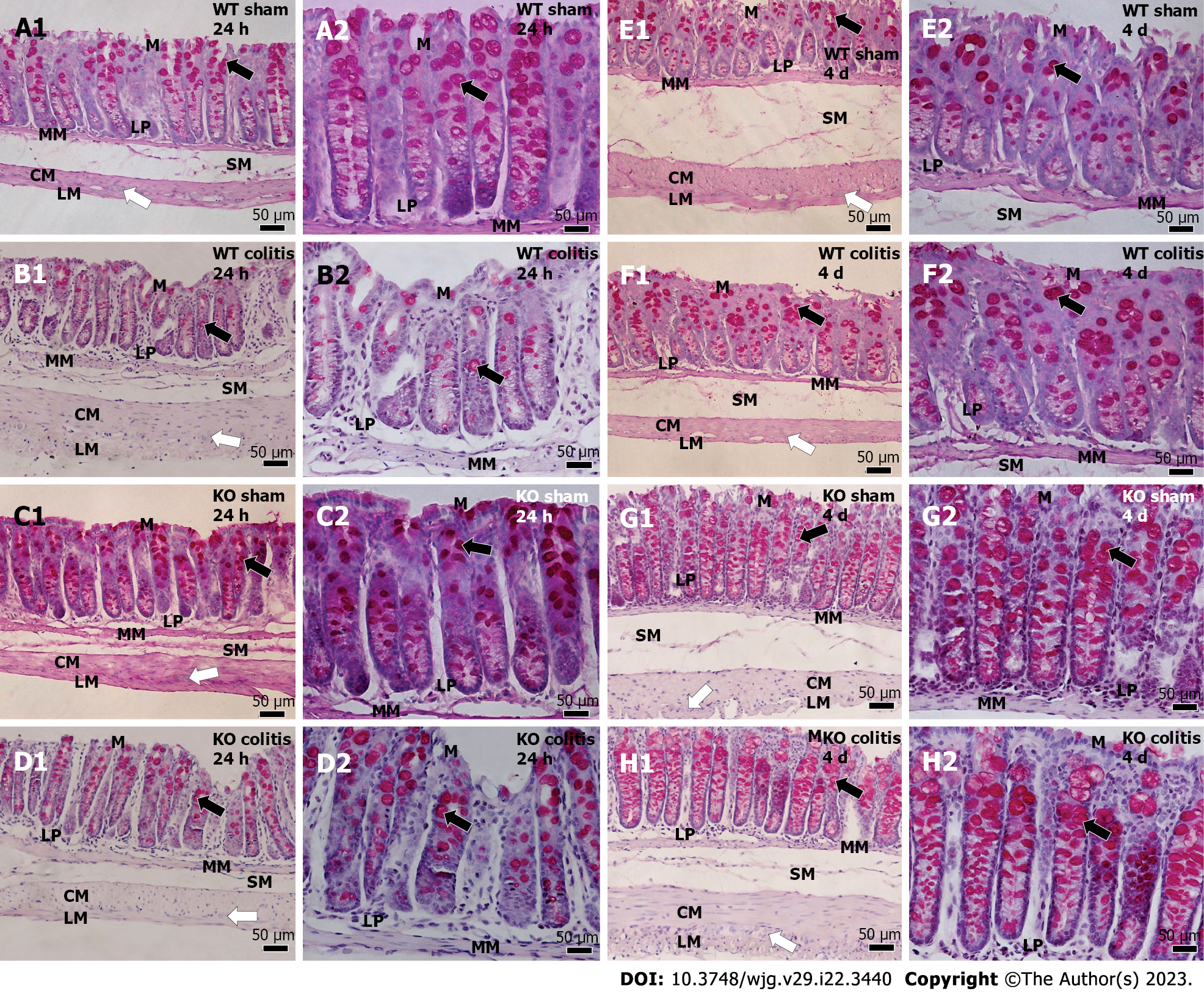
Figure 15 Photomicrograph of periodic acid Schiff-stained transverse sections of the distal colons of mice.
A1 and A2: Wild-type (WT) sham 24 h; B1 and B2: WT colitis 24 h; C1 and C2: Knockout (KO) sham 24 h; D1 and D2: KO colitis 24 h; E1 and E2: WT sham 4 d; F1 and F2: WT colitis 4 d; G1 and G2: KO sham 4 d; H1 and H2: KO colitis 4 d groups. Goblet cells were observed throughout the epithelium in the sham groups, and there was an apparent reduction in goblet cell number and size in the WT colitis group. In addition, goblet cells showed a more rounded shape and an intumescent appearance in the WT colitis group. In the KO groups, an apparent increase in the number of goblet cells was noted. WT: Wild-type; KO: Knockout; M: Mucosal layer; LP: Lamina propria; MM: Mucosal muscle; SM: Submucosal layer; ML: Muscle layer; CM: Circular musculature; LM: Longitudinal musculature; white arrows: Myenteric ganglion; black arrows: Goblet cells.
- Citation: Magalhães HIR, Machado FA, Souza RF, Caetano MAF, Figliuolo VR, Coutinho-Silva R, Castelucci P. Study of the roles of caspase-3 and nuclear factor kappa B in myenteric neurons in a P2X7 receptor knockout mouse model of ulcerative colitis. World J Gastroenterol 2023; 29(22): 3440-3468
- URL: https://www.wjgnet.com/1007-9327/full/v29/i22/3440.htm
- DOI: https://dx.doi.org/10.3748/wjg.v29.i22.3440


