Copyright
©The Author(s) 2023.
World J Gastroenterol. Jun 14, 2023; 29(22): 3440-3468
Published online Jun 14, 2023. doi: 10.3748/wjg.v29.i22.3440
Published online Jun 14, 2023. doi: 10.3748/wjg.v29.i22.3440
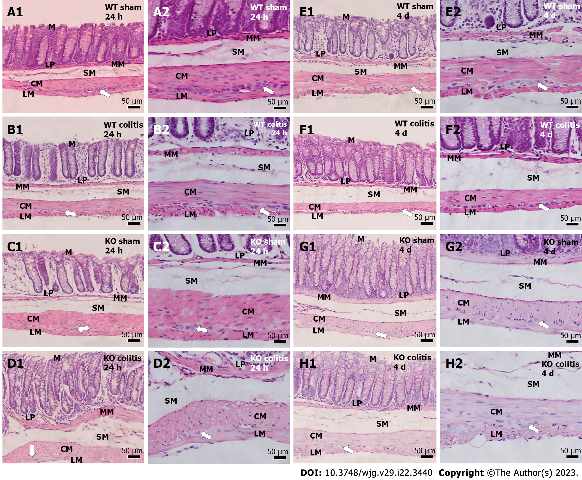
Figure 12 Photomicrographs of hematoxylin-eosin-stained transverse sections of the distal colons of mice of different groups.
A1 and A2: Wild-type (WT) sham 24 h; B1 and B2: WT colitis 24 h; C1 and C2: Knockout (KO) sham 24 h; D1 and D2: KO colitis 24 h; E1 and E2: WT sham 4 d; F1 and F2: WT colitis 4 d; G1 and G2: KO sham 4 d; H1 and H2: KO colitis 4 d groups. Tissue integrity was maintained in the sham groups, and cellular disruption and thickening of the lamina propria and submucosal layer were observed in the WT colitis group. Tissue was better preserved, and there were fewer inflammatory cells in the KO colitis group than in the WT colitis group. M: Mucosal layer; LP: Lamina propria; MM: Mucosal muscle; SM: Submucosal layer; ML: Muscle layer; CM: Circular musculature; LM: Longitudinal musculature; WT: Wild-type; KO: Knockout. White arrows mean myenteric ganglion.
- Citation: Magalhães HIR, Machado FA, Souza RF, Caetano MAF, Figliuolo VR, Coutinho-Silva R, Castelucci P. Study of the roles of caspase-3 and nuclear factor kappa B in myenteric neurons in a P2X7 receptor knockout mouse model of ulcerative colitis. World J Gastroenterol 2023; 29(22): 3440-3468
- URL: https://www.wjgnet.com/1007-9327/full/v29/i22/3440.htm
- DOI: https://dx.doi.org/10.3748/wjg.v29.i22.3440


